 Movie
Movie Controller

[English] 日本語
 Yorodumi
- PDB-2du0: Crystal structure of basic winged bean lectin in complex with Alp... -
+ Open data

- Basic information
| Entry | Database: PDB / ID: 2du0 | |||||||||
|---|---|---|---|---|---|---|---|---|---|---|
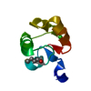
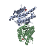
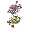
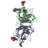
| Title | Crystal structure of basic winged bean lectin in complex with Alpha-D-galactose | |||||||||
 Components Components | Basic agglutinin | |||||||||
 Keywords Keywords | SUGAR BINDING PROTEIN /  LEGUME LECTIN / GLYCOSYLATED PROTEIN / AGGLUTININ LEGUME LECTIN / GLYCOSYLATED PROTEIN / AGGLUTININ | |||||||||
| Function / homology |  Function and homology information Function and homology information | |||||||||
| Biological species |   Psophocarpus tetragonolobus (winged bean) Psophocarpus tetragonolobus (winged bean) | |||||||||
| Method | X-RAY DIFFRACTION / Resolution: 2.7 Å | |||||||||
 Authors Authors | Kulkarni, K.A. / Katiyar, S. / Surolia, A. / Vijayan, M. / Suguna, K. | |||||||||
 Citation Citation | Journal: ACTA CRYSTALLOGR.,SECT.D / Year: 2006 Title: Structural basis for the carbohydrate-specificity of basic winged-bean lectin and its differential affinity for Gal and GalNAc Authors: Kulkarni, K.A. / Katiyar, S. / Surolia, A. / Vijayan, M. / Suguna, K. #1: Journal: J.Mol.Biol. / Year: 1998Title: Carbohydrate specificity and quaternary association in basic winged bean lectin: X-ray analysis of the lectin at 2.5 A resolution Authors: Prabu, M.M. / Sankaranarayanan, R. / Puri, K.D. / Sharma, V. / Surolia, A. / Vijayan, M. / Suguna, K. #2: Journal: Febs Lett. / Year: 2005Title: Structural basis for the specificity of basic winged bean lectin for the Tn-antigen: a crystallographic, thermodynamic and modelling study Authors: Kulkarni, K.A. / Surolia, A. / Vijayan, M. / Suguna, K. #3: Journal: ACTA CRYSTALLOGR.,SECT.D / Year: 1999Title: Structure of basic winged-bean lectin and a comparison with its saccharide-bound form Authors: Manoj, N. / Srinivas, V.R. / Surolia, A. / Vijayan, M. / Suguna, K. | |||||||||
| History |
|
- Structure visualization
| Structure viewer | Molecule: MolmilJmol/JSmol |
|---|
- Downloads & links
-Download
| PDBx/mmCIF format | 2du0.cif.gz | 204.2 KB | Display | PDBx/mmCIF format |
|---|---|---|---|---|
| PDB format | pdb2du0.ent.gz | 162.6 KB | Display | PDB format |
| PDBx/mmJSON format | 2du0.json.gz | Tree view | PDBx/mmJSON format | |
| Others |  Other downloads Other downloads |
-Validation report
| Arichive directory | https://data.pdbj.org/pub/pdb/validation_reports/du/2du0ftp://data.pdbj.org/pub/pdb/validation_reports/du/2du0 | HTTPS FTP |
|---|
-Related structure data
| Related structure data |  2dtwC  2dtyC  2du1C  1wblS S: Starting model for refinement C: citing same article ( |
|---|---|
| Similar structure data |


-Links
 PDBj
PDBj

- Assembly
| Deposited unit | 
| ||||||||
|---|---|---|---|---|---|---|---|---|---|
| 1 | 
| ||||||||
| 2 | 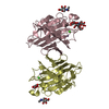
| ||||||||
| Unit cell |
| ||||||||
| Components on special symmetry positions |
| ||||||||
| Details | It is a homodimer formed by the subunits A&B and C&D. |
-Components
-Protein , 1 types, 4 molecules ABCD
| #1: Protein | Mass: 26505.604 Da / Num. of mol.: 4 / Source method: isolated from a natural source / Source: (natural) Psophocarpus tetragonolobus (winged bean) / References: UniProt: O24313 |
|---|
-Sugars , 4 types, 11 molecules 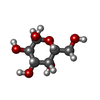

| #2: Polysaccharide | / Mass: 570.542 Da / Num. of mol.: 3 Source method: isolated from a genetically manipulated source #3: Polysaccharide | alpha-L-fucopyranose-(1-3)-2-acetamido-2-deoxy-beta-D-glucopyranose | / Mass: 367.349 Da / Num. of mol.: 1Source method: isolated from a genetically manipulated source #4: Sugar | ChemComp-GLA / Galactose Type: D-saccharide, alpha linking / Mass: 180.156 Da / Num. of mol.: 4 Type: D-saccharide, alpha linking / Mass: 180.156 Da / Num. of mol.: 4Source method: isolated from a genetically manipulated source Formula: C6H12O6 #5: Sugar | N-Acetylglucosamine Type: D-saccharide, beta linking / Mass: 221.208 Da / Num. of mol.: 3 Type: D-saccharide, beta linking / Mass: 221.208 Da / Num. of mol.: 3Source method: isolated from a genetically manipulated source Formula: C8H15NO6 |
|---|
-Non-polymers , 3 types, 247 molecules 


| #6: Chemical | ChemComp-CA /  Mass: 40.078 Da / Num. of mol.: 4 / Source method: obtained synthetically / Formula: Ca Mass: 40.078 Da / Num. of mol.: 4 / Source method: obtained synthetically / Formula: Ca#7: Chemical | ChemComp-MN /  Mass: 54.938 Da / Num. of mol.: 4 / Source method: obtained synthetically / Formula: Mn Mass: 54.938 Da / Num. of mol.: 4 / Source method: obtained synthetically / Formula: Mn#8: Water | ChemComp-HOH / | WaterMass: 18.015 Da / Num. of mol.: 239 / Source method: isolated from a natural source / Formula: H2O |
|---|
-Experimental details
-Experiment
| Experiment | Method: X-RAY DIFFRACTION / Number of used crystals: 1 |
|---|
- Sample preparation
| Crystal | Density Matthews: 2.45 Å3/Da / Density % sol: 49.83 % |
|---|---|
| Crystal grow | Temperature: 300 K / Method: vapor diffusion, hanging drop / pH: 7.4 Details: 20% PEG 4000, 10% Iso-propenol, 0.02M PBS, pH 7.4, VAPOR DIFFUSION, HANGING DROP, temperature 300K |
-Data collection
| Diffraction | Mean temperature: 298 K |
|---|---|
| Diffraction source | Source: ROTATING ANODE / Type: RIGAKU RU300 / Wavelength: 1.5418 Å |
| Detector | Type: MAR scanner 345 mm plate / Detector: IMAGE PLATE |
| Radiation | Monochromator: Osmic Mirrors / Protocol: SINGLE WAVELENGTH / Monochromatic (M) / Laue (L): M / Scattering type: x-ray |
| Radiation wavelength | Wavelength: 1.5418 Å / Relative weight: 1 |
| Reflection | Resolution: 2.7→30 Å / Num. obs: 29164 / % possible obs: 99.2 % / Observed criterion σ(F): 1 / Observed criterion σ(I): 1 / Biso Wilson estimate: 52.6 Å2 / Rmerge(I) obs: 0.114 / Net I/σ(I): 16.8 |
| Reflection shell | Resolution: 2.7→2.8 Å / Rmerge(I) obs: 0.316 / Mean I/σ(I) obs: 4.2 / Num. unique all: 2865 / % possible all: 99.3 |
- Processing
| Software | Name: CNS / Version: 1.1 / Classification: refinement | ||||||||||||||||||||||||||||||||||||
|---|---|---|---|---|---|---|---|---|---|---|---|---|---|---|---|---|---|---|---|---|---|---|---|---|---|---|---|---|---|---|---|---|---|---|---|---|---|
| Refinement | Starting model: PDB entry 1wbl Resolution: 2.7→30 Å / Rfactor Rfree error: 0.007 / Data cutoff high absF: 2172589.67 / Data cutoff low absF: 0 / Isotropic thermal model: RESTRAINED / Cross valid method: THROUGHOUT / σ(F): 0
| ||||||||||||||||||||||||||||||||||||
| Solvent computation | Solvent model: FLAT MODEL / Bsol: 36.4292 Å2 / ksol: 0.288649 e/Å3 | ||||||||||||||||||||||||||||||||||||
| Displacement parameters | Biso mean: 38.8 Å2
| ||||||||||||||||||||||||||||||||||||
| Refine analyze |
| ||||||||||||||||||||||||||||||||||||
| Refinement step | Cycle: LAST / Resolution: 2.7→30 Å
| ||||||||||||||||||||||||||||||||||||
| Refine LS restraints |
| ||||||||||||||||||||||||||||||||||||
| LS refinement shell | Resolution: 2.7→2.8 Å / Rfactor Rfree error: 0.034 / Total num. of bins used: 10
| ||||||||||||||||||||||||||||||||||||
| Xplor file |
|
