 Movie
Movie Controller

[English] 日本語
 Yorodumi
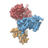
- EMDB-26426: Meprin alpha helix in complex with fetuin-B [subparticle localise... -
+ Open data

- Basic information
| Entry |  | |||||||||
|---|---|---|---|---|---|---|---|---|---|---|
| Title | Meprin alpha helix in complex with fetuin-B [subparticle localised reconstruction, masked focused refinement] | |||||||||
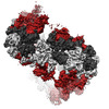
 Map data Map data | B-factor amplitude corrected sharpened map, filtered by local resolution | |||||||||
 Sample Sample |
| |||||||||
| Function / homology |  Function and homology information Function and homology information meprin A / meprin A complex / epidermal growth factor receptor ligand maturation / metalloendopeptidase inhibitor activity / metallodipeptidase activity / negative regulation of endopeptidase activity / binding of sperm to zona pellucida / cysteine-type endopeptidase inhibitor activity / endopeptidase inhibitor activity / signaling receptor ligand precursor processing ...meprin A / meprin A complex / epidermal growth factor receptor ligand maturation / metalloendopeptidase inhibitor activity / metallodipeptidase activity / negative regulation of endopeptidase activity / binding of sperm to zona pellucida / cysteine-type endopeptidase inhibitor activity / endopeptidase inhibitor activity / signaling receptor ligand precursor processing / single fertilization / metalloendopeptidase activity / metallopeptidase activity / extracellular space / extracellular exosome / zinc ion binding / extracellular region / plasma membrane meprin A / meprin A complex / epidermal growth factor receptor ligand maturation / metalloendopeptidase inhibitor activity / metallodipeptidase activity / negative regulation of endopeptidase activity / binding of sperm to zona pellucida / cysteine-type endopeptidase inhibitor activity / endopeptidase inhibitor activity / signaling receptor ligand precursor processing ...meprin A / meprin A complex / epidermal growth factor receptor ligand maturation / metalloendopeptidase inhibitor activity / metallodipeptidase activity / negative regulation of endopeptidase activity / binding of sperm to zona pellucida / cysteine-type endopeptidase inhibitor activity / endopeptidase inhibitor activity / signaling receptor ligand precursor processing / single fertilization / metalloendopeptidase activity / metallopeptidase activity / extracellular space / extracellular exosome / zinc ion binding / extracellular region / plasma membraneSimilarity search - Function | |||||||||
| Biological species |  Homo sapiens (human) Homo sapiens (human) | |||||||||
| Method | single particle reconstruction / cryo EM / Resolution: 2.8 Å | |||||||||
 Authors Authors | Bayly-Jones C / Lupton CJ / Fritz C / Schlenzig D / Whisstock JC | |||||||||
| Funding support |  Germany, Germany,  Australia, 2 items Australia, 2 items
| |||||||||
 Citation Citation | Journal: Nat Commun / Year: 2022 Title: Helical ultrastructure of the metalloprotease meprin α in complex with a small molecule inhibitor. Authors: Charles Bayly-Jones / Christopher J Lupton / Claudia Fritz / Hariprasad Venugopal / Daniel Ramsbeck / Michael Wermann / Christian Jäger / Alex de Marco / Stephan Schilling / Dagmar ...Authors: Charles Bayly-Jones / Christopher J Lupton / Claudia Fritz / Hariprasad Venugopal / Daniel Ramsbeck / Michael Wermann / Christian Jäger / Alex de Marco / Stephan Schilling / Dagmar Schlenzig / James C Whisstock / Abstract: The zinc-dependent metalloprotease meprin α is predominantly expressed in the brush border membrane of proximal tubules in the kidney and enterocytes in the small intestine and colon. In normal ...The zinc-dependent metalloprotease meprin α is predominantly expressed in the brush border membrane of proximal tubules in the kidney and enterocytes in the small intestine and colon. In normal tissue homeostasis meprin α performs key roles in inflammation, immunity, and extracellular matrix remodelling. Dysregulated meprin α is associated with acute kidney injury, sepsis, urinary tract infection, metastatic colorectal carcinoma, and inflammatory bowel disease. Accordingly, meprin α is the target of drug discovery programs. In contrast to meprin β, meprin α is secreted into the extracellular space, whereupon it oligomerises to form giant assemblies and is the largest extracellular protease identified to date (~6 MDa). Here, using cryo-electron microscopy, we determine the high-resolution structure of the zymogen and mature form of meprin α, as well as the structure of the active form in complex with a prototype small molecule inhibitor and human fetuin-B. Our data reveal that meprin α forms a giant, flexible, left-handed helical assembly of roughly 22 nm in diameter. We find that oligomerisation improves proteolytic and thermal stability but does not impact substrate specificity or enzymatic activity. Furthermore, structural comparison with meprin β reveal unique features of the active site of meprin α, and helical assembly more broadly. | |||||||||
| History |
|
- Structure visualization
| Supplemental images |
|---|

- Downloads & links
-EMDB archive
| Map data | emd_26426.map.gz | 1.6 MB | EMDB map data format | |
|---|---|---|---|---|
| Header (meta data) | emd-26426-v30.xmlemd-26426.xml | 29.1 KB 29.1 KB | Display Display | EMDB header |
| FSC (resolution estimation) | emd_26426_fsc.xml | 7 KB | Display | FSC data file |
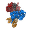
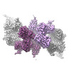
| Images | 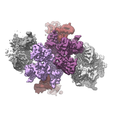 emd_26426.png emd_26426.png | 104 KB | ||
| Masks | emd_26426_msk_1.map | 38.4 MB | Mask map | |
| Others | emd_26426_additional_1.map.gzemd_26426_additional_2.map.gzemd_26426_half_map_1.map.gzemd_26426_half_map_2.map.gz | 34.2 MB 33.1 MB 35.7 MB 35.7 MB | ||
| Archive directory |  http://ftp.pdbj.org/pub/emdb/structures/EMD-26426ftp://ftp.pdbj.org/pub/emdb/structures/EMD-26426 http://ftp.pdbj.org/pub/emdb/structures/EMD-26426ftp://ftp.pdbj.org/pub/emdb/structures/EMD-26426 | HTTPS FTP |
-Related structure data
| Related structure data | 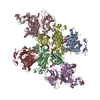 7uaiMC  7uabC  7uacC  7uaeC 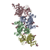 7uafC C: citing same article ( M: atomic model generated by this map |
|---|---|
| Similar structure data |
-Links
| EMDB pages | EMDB (EBI/PDBe) / EMDataResource |
|---|---|
| Related items in Molecule of the Month |


-Map
| File | Download / File: emd_26426.map.gz / Format: CCP4 / Size: 38.4 MB / Type: IMAGE STORED AS FLOATING POINT NUMBER (4 BYTES) | ||||||||||||||||||||
|---|---|---|---|---|---|---|---|---|---|---|---|---|---|---|---|---|---|---|---|---|---|
| Annotation | B-factor amplitude corrected sharpened map, filtered by local resolution | ||||||||||||||||||||
| Voxel size | X=Y=Z: 1.41333 Å | ||||||||||||||||||||
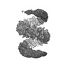
| Density |
| ||||||||||||||||||||
| Symmetry | Space group: 1 | ||||||||||||||||||||
| Details | EMDB XML:
|
-Supplemental data
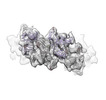
-Mask #1
| File | emd_26426_msk_1.map | ||||||||||||
|---|---|---|---|---|---|---|---|---|---|---|---|---|---|
| Projections & Slices |
| ||||||||||||
| Density Histograms |
 Z
Z Y
Y X
X


-Additional map: DeepEMhancer (mask) sharpened final map. Low pass filtered...
| File | emd_26426_additional_1.map | ||||||||||||
|---|---|---|---|---|---|---|---|---|---|---|---|---|---|
| Annotation | DeepEMhancer (mask) sharpened final map. Low pass filtered to global FSC. | ||||||||||||
| Projections & Slices |
| ||||||||||||
| Density Histograms |


-Additional map: DeepEMhancer (highres) sharpened final map. Low pass filtered...
| File | emd_26426_additional_2.map | ||||||||||||
|---|---|---|---|---|---|---|---|---|---|---|---|---|---|
| Annotation | DeepEMhancer (highres) sharpened final map. Low pass filtered to global FSC. | ||||||||||||
| Projections & Slices |
| ||||||||||||
| Density Histograms |


-Half map: Unfiltered half-map (1 of 2).
| File | emd_26426_half_map_1.map | ||||||||||||
|---|---|---|---|---|---|---|---|---|---|---|---|---|---|
| Annotation | Unfiltered half-map (1 of 2). | ||||||||||||
| Projections & Slices |
| ||||||||||||
| Density Histograms |


-Half map: Unfiltered half-map (2 of 2).
| File | emd_26426_half_map_2.map | ||||||||||||
|---|---|---|---|---|---|---|---|---|---|---|---|---|---|
| Annotation | Unfiltered half-map (2 of 2). | ||||||||||||
| Projections & Slices |
| ||||||||||||
| Density Histograms |


- Sample components
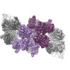
-Entire : Tetrameric portion of meprin alpha helix in complex with fetuin-B...
| Entire | Name: Tetrameric portion of meprin alpha helix in complex with fetuin-B [subparticle localised reconstruction] |
|---|---|
| Components |
|
-Supramolecule #1: Tetrameric portion of meprin alpha helix in complex with fetuin-B...
| Supramolecule | Name: Tetrameric portion of meprin alpha helix in complex with fetuin-B [subparticle localised reconstruction] type: complex / Chimera: Yes / ID: 1 / Parent: 0 / Macromolecule list: #1-#2 Details: Subparticle localised reconstruction of tetrameric region of recombinant, secreted helical meprin alpha in complex with fetuin-B |
|---|---|
| Source (natural) | Organism: Homo sapiens (human) |
| Recombinant expression | Organism:  Drosophila (fruit flies) / Recombinant cell: Schneider-2 / Recombinant plasmid: pMT/BiP/V5 Drosophila (fruit flies) / Recombinant cell: Schneider-2 / Recombinant plasmid: pMT/BiP/V5 |
| Molecular weight | Theoretical: 85 kDa/nm |
-Macromolecule #1: Meprin A subunit alpha
| Macromolecule | Name: Meprin A subunit alpha / type: protein_or_peptide / ID: 1 / Number of copies: 4 / Enantiomer: LEVO / EC number: meprin A |
|---|---|
| Source (natural) | Organism: Homo sapiens (human) |
| Molecular weight | Theoretical: 67.28582 KDa |
| Recombinant expression | Organism: Drosophila melanogaster (fruit fly) |
| Sequence | String: WSHPQFEKVP IKYLPEENVH DADFGEQKDI SEINLAAGLD LFQGDILLQK SRNGLRDPNT RWTFPIPYIL ADNLGLNAKG AILYAFEMF RLKSCVDFKP YEGESSYIIF QQFDGCWSEV GDQHVGQNIS IGQGCAYKAI IEHEILHALG FYHEQSRTDR D DYVNIWWD ...String: WSHPQFEKVP IKYLPEENVH DADFGEQKDI SEINLAAGLD LFQGDILLQK SRNGLRDPNT RWTFPIPYIL ADNLGLNAKG AILYAFEMF RLKSCVDFKP YEGESSYIIF QQFDGCWSEV GDQHVGQNIS IGQGCAYKAI IEHEILHALG FYHEQSRTDR D DYVNIWWD QILSGYQHNF DTYDDSLITD LNTPYDYESL MHYQPFSFNK NASVPTITAK IPEFNSIIGQ RLDFSAIDLE RL NRMYNCT TTHTLLDHCT FEKANICGMI QGTRDDTDWA HQDSAQAGEV DHTLLGQCTG AGYFMQFSTS SGSAEEAALL ESR ILYPKR KQQCLQFFYK MTGSPSDRLV VWVRRDDSTG NVRKLVKVQT FQGDDDHNWK IAHVVLKEEQ KFRYLFQGTK GDPQ NSTGG IYLDDITLTE TPCPTGVWTV RNFSQVLENT SKGDKLQSPR FYNSEGYGFG VTLYPNSRES SGYLRLAFHV CSGEN DAIL EWPVENRQVI ITILDQEPDV RNRMSSSMVF TTSKSHTSPA INDTVIWDRP SRVGTYHTDC NCFRSIDLGW SGFISH QML KRRSFLKNDD LIIFVDFEDI THLS |
-Macromolecule #2: Fetuin-B
| Macromolecule | Name: Fetuin-B / type: protein_or_peptide / ID: 2 / Number of copies: 2 / Enantiomer: LEVO |
|---|---|
| Source (natural) | Organism: Homo sapiens (human) |
| Molecular weight | Theoretical: 42.926691 KDa |
| Recombinant expression | Organism: Homo sapiens (human) |
| Sequence | String: MGLLLPLALC ILVLCCGAMS PPQLALNPSA LLSRGCNDSD VLAVAGFALR DINKDRKDGY VLRLNRVNDA QEYRRGGLGS LFYLTLDVL ETDCHVLRKK AWQDCGMRIF FESVYGQCKA IFYMNNPSRV LYLAAYNCTL RPVSKKKIYM TCPDCPSSIP T DSSNHQVL ...String: MGLLLPLALC ILVLCCGAMS PPQLALNPSA LLSRGCNDSD VLAVAGFALR DINKDRKDGY VLRLNRVNDA QEYRRGGLGS LFYLTLDVL ETDCHVLRKK AWQDCGMRIF FESVYGQCKA IFYMNNPSRV LYLAAYNCTL RPVSKKKIYM TCPDCPSSIP T DSSNHQVL EAATESLAKY NNENTSKQYS LFKVTRASSQ WVVGPSYFVE YLIKESPCTK SQASSCSLQS SDSVPVGLCK GS LTRTHWE KFVSVTCDFF ESQAPATGSE NSAVNQKPTN LPKVEESQQK NTPPTDSPSK AGPRGSVQYL PDLDDKNSQE KGP QEAFPV HLDLTTNPQG ETLDISFLFL EPMEEKLVVL PFPKEKARTA ECPGPAQNAS PLVLPPHHHH HH |
-Macromolecule #6: CALCIUM ION
| Macromolecule | Name: CALCIUM ION / type: ligand / ID: 6 / Number of copies: 8 / Formula: CA |
|---|---|
| Molecular weight | Theoretical: 40.078 Da |
-Macromolecule #7: ZINC ION
| Macromolecule | Name: ZINC ION / type: ligand / ID: 7 / Number of copies: 4 / Formula: ZN |
|---|---|
| Molecular weight | Theoretical: 65.409 Da |
-Macromolecule #8: 2-acetamido-2-deoxy-beta-D-glucopyranose
| Macromolecule | Name: 2-acetamido-2-deoxy-beta-D-glucopyranose / type: ligand / ID: 8 / Number of copies: 4 / Formula: NAG |
|---|---|
| Molecular weight | Theoretical: 221.208 Da |
| Chemical component information |  ChemComp-NAG: |
-Experimental details
-Structure determination
| Method | cryo EM |
|---|---|
 Processing Processing | single particle reconstruction |
| Aggregation state | filament |
-Sample preparation
| Concentration | 1.0 mg/mL | |||||||||
|---|---|---|---|---|---|---|---|---|---|---|
| Buffer | pH: 7.4 Component:
| |||||||||
| Grid | Model: Quantifoil R2/2 / Material: COPPER / Mesh: 200 / Support film - Material: CARBON / Support film - topology: HOLEY / Pretreatment - Type: GLOW DISCHARGE / Pretreatment - Atmosphere: AIR / Details: Pelco EasyGlow | |||||||||
| Vitrification | Cryogen name: ETHANE / Chamber humidity: 100 % / Chamber temperature: 277.15 K / Instrument: FEI VITROBOT MARK IV / Details: 3 s blot, -3 force. |
- Electron microscopy
| Microscope | FEI TITAN KRIOS |
|---|---|
| Electron beam | Acceleration voltage: 300 kV / Electron source: FIELD EMISSION GUN |
| Electron optics | Illumination mode: FLOOD BEAM / Imaging mode: BRIGHT FIELDBright-field microscopy / Cs: 2.7 mm / Nominal defocus max: 2.0 µm / Nominal defocus min: 0.5 µm / Nominal magnification: 130000 |
| Specialist optics | Energy filter - Name: GIF Bioquantum |
| Sample stage | Specimen holder model: FEI TITAN KRIOS AUTOGRID HOLDER / Cooling holder cryogen: NITROGEN |
| Image recording | Film or detector model: GATAN K3 (6k x 4k) / Detector mode: COUNTING / Digitization - Frames/image: 1-25 / Number grids imaged: 1 / Number real images: 4068 / Average electron dose: 44.5 e/Å2 |
| Experimental equipment |  Model: Titan Krios / Image courtesy: FEI Company |
-Image processing
| Particle selection | Number selected: 519760 Details: Localised subparticles extrated after expansion of pseudo-helical symmetry | ||||||
|---|---|---|---|---|---|---|---|
| CTF correction | Software: (Name: CTFFIND (ver. 4.1.13), Warp (ver. 1.0.7)) | ||||||
| Startup model | Type of model: OTHER / Details: Ab initio volume | ||||||
| Initial angle assignment | Type: MAXIMUM LIKELIHOOD Software:
| ||||||
| Final 3D classification | Number classes: 8 / Avg.num./class: 100000 / Software - Name: RELION (ver. 3.1) / Details: Focused classification without changes to angles | ||||||
| Final angle assignment | Type: MAXIMUM LIKELIHOOD Software:
| ||||||
| Final reconstruction | Applied symmetry - Point group: C1 (asymmetric) / Algorithm: FOURIER SPACE / Resolution.type: BY AUTHOR / Resolution: 2.8 Å / Resolution method: FSC 0.143 CUT-OFF / Number images used: 115951 | ||||||
| Details | Compressed to LZW TIFF. Motion corrected by MotionCor. | ||||||
| FSC plot (resolution estimation) |  |

