 Movie
Movie Controller
 Structure viewers
 About Yorodumi Papers


+Search query
-Structure paper
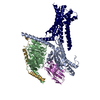
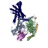
| Title | Molecular features of the ligand-free GLP-1R, GCGR and GIPR in complex with G proteins. |
|---|---|
| Journal, issue, pages | Cell Discov, Vol. 10, Issue 1, Page 18, Year 2024 |
| Publish date | Feb 13, 2024 |
 Authors Authors | Zhaotong Cong / Fenghui Zhao / Yang Li / Gan Luo / Yiting Mai / Xianyue Chen / Yanyan Chen / Shi Lin / Xiaoqing Cai / Qingtong Zhou / Dehua Yang / Ming-Wei Wang /   |
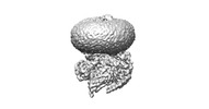
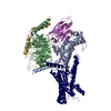
| PubMed Abstract | Class B1 G protein-coupled receptors (GPCRs) are important regulators of many physiological functions such as glucose homeostasis, which is mainly mediated by three peptide hormones, i.e., glucagon- ...Class B1 G protein-coupled receptors (GPCRs) are important regulators of many physiological functions such as glucose homeostasis, which is mainly mediated by three peptide hormones, i.e., glucagon-like peptide-1 (GLP-1), glucagon (GCG), and glucose-dependent insulinotropic polypeptide (GIP). They trigger a cascade of signaling events leading to the formation of an active agonist-receptor-G protein complex. However, intracellular signal transducers can also activate the receptor independent of extracellular stimuli, suggesting an intrinsic role of G proteins in this process. Here, we report cryo-electron microscopy structures of the human GLP-1 receptor (GLP-1R), GCG receptor (GCGR), and GIP receptor (GIPR) in complex with G proteins without the presence of cognate ligands. These ligand-free complexes share a similar intracellular architecture to those bound by endogenous peptides, in which, the G protein alone directly opens the intracellular binding cavity and rewires the extracellular orthosteric pocket to stabilize the receptor in a state unseen before. While the peptide-binding site is partially occupied by the inward folded transmembrane helix 6 (TM6)-extracellular loop 3 (ECL3) juncture of GIPR or a segment of GCGR ECL2, the extracellular portion of GLP-1R adopts a conformation close to the active state. Our findings offer valuable insights into the distinct activation mechanisms of these three important receptors. It is possible that in the absence of a ligand, the intracellular half of transmembrane domain is mobilized with the help of G protein, which in turn rearranges the extracellular half to form a transitional conformation, facilitating the entry of the peptide N-terminus. |
 External links External links | Cell Discov / PubMed:38346960 / PubMed Central |
| Methods | EM (single particle) |
| Resolution | 2.54 - 2.86 Å |
| Structure data | EMDB-37390, PDB-8wa3: EMDB-37504, PDB-8wg7: EMDB-37505, PDB-8wg8: |
| Source |
|
 Keywords Keywords |  STRUCTURAL PROTEIN / Glucose-dependent insulinotropic polypeptide receptor; cryo-electron microscopy; G protein-coupled receptor; ligand recognition / MEMBRANE PROTEIN / G protein-coupled receptor / ligand recognition / receptor activation / unimolecular agonist STRUCTURAL PROTEIN / Glucose-dependent insulinotropic polypeptide receptor; cryo-electron microscopy; G protein-coupled receptor; ligand recognition / MEMBRANE PROTEIN / G protein-coupled receptor / ligand recognition / receptor activation / unimolecular agonist |


 leptolinea tardivitalis (bacteria)

