Movie
Movie Controller


+ Open data

- Basic information
| Entry |  | |||||||||||||||
|---|---|---|---|---|---|---|---|---|---|---|---|---|---|---|---|---|
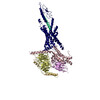
| Title | Cryo-EM structure of peptide free and Gs-coupled GIPR | |||||||||||||||
 Map data Map data | ||||||||||||||||
 Sample Sample |
| |||||||||||||||
 Keywords Keywords | Glucose-dependent insulinotropic polypeptide receptor /  cryo-electron microscopy / G protein-coupled receptor / ligand recognition / STRUCTURAL PROTEIN cryo-electron microscopy / G protein-coupled receptor / ligand recognition / STRUCTURAL PROTEIN | |||||||||||||||
| Function / homology |  Function and homology information Function and homology informationgastric inhibitory peptide receptor activity / glucagon family peptide binding / gastric inhibitory peptide signaling pathway / desensitization of G protein-coupled receptor signaling pathway / sensory perception of chemical stimulus / endocrine pancreas development / mu-type opioid receptor binding / response to fatty acid / corticotropin-releasing hormone receptor 1 binding / G-protein activation ...gastric inhibitory peptide receptor activity / glucagon family peptide binding / gastric inhibitory peptide signaling pathway / desensitization of G protein-coupled receptor signaling pathway / sensory perception of chemical stimulus / endocrine pancreas development / mu-type opioid receptor binding / response to fatty acid / corticotropin-releasing hormone receptor 1 binding / G-protein activation / Activation of the phototransduction cascade / : / Glucagon-type ligand receptors / Thromboxane signalling through TP receptor / Sensory perception of sweet, bitter, and umami (glutamate) taste / G beta:gamma signalling through PI3Kgamma / G beta:gamma signalling through CDC42 / Cooperation of PDCL (PhLP1) and TRiC/CCT in G-protein beta folding / Ca2+ pathway / Activation of G protein gated Potassium channels / Synthesis, secretion, and inactivation of Glucagon-like Peptide-1 (GLP-1) / Inhibition of voltage gated Ca2+ channels via Gbeta/gamma subunits / G protein-coupled peptide receptor activity / G alpha (z) signalling events / Vasopressin regulates renal water homeostasis via Aquaporins / Glucagon-like Peptide-1 (GLP1) regulates insulin secretion / Adrenaline,noradrenaline inhibits insulin secretion / ADP signalling through P2Y purinoceptor 12 / G alpha (q) signalling events / G alpha (i) signalling events / Thrombin signalling through proteinase activated receptors (PARs) / Activation of G protein gated Potassium channels / G-protein activation / G beta:gamma signalling through PI3Kgamma / Prostacyclin signalling through prostacyclin receptor / G beta:gamma signalling through PLC beta / ADP signalling through P2Y purinoceptor 1 / Thromboxane signalling through TP receptor / Presynaptic function of Kainate receptors / G beta:gamma signalling through CDC42 / Inhibition of voltage gated Ca2+ channels via Gbeta/gamma subunits / Glucagon-type ligand receptors / alkylglycerophosphoethanolamine phosphodiesterase activity / Adrenaline,noradrenaline inhibits insulin secretion / G alpha (12/13) signalling events / G beta:gamma signalling through BTK / ADP signalling through P2Y purinoceptor 12 / Cooperation of PDCL (PhLP1) and TRiC/CCT in G-protein beta folding / Thrombin signalling through proteinase activated receptors (PARs) / Ca2+ pathway / G alpha (z) signalling events / Extra-nuclear estrogen signaling / G alpha (s) signalling events / beta-2 adrenergic receptor binding / G alpha (q) signalling events / photoreceptor outer segment membrane / G alpha (i) signalling events / Glucagon-like Peptide-1 (GLP1) regulates insulin secretion / spectrin binding / Vasopressin regulates renal water homeostasis via Aquaporins / peptide hormone binding / regulation of insulin secretion / photoreceptor outer segment / D1 dopamine receptor binding / response to axon injury / positive regulation of cAMP-mediated signaling / response to glucose / adenylate cyclase-activating adrenergic receptor signaling pathway / cardiac muscle cell apoptotic process / ionotropic glutamate receptor binding / insulin-like growth factor receptor binding / activation of adenylate cyclase activity / photoreceptor inner segment / adenylate cyclase activator activity / response to nutrient / generation of precursor metabolites and energy / G-protein beta/gamma-subunit complex binding / adenylate cyclase-modulating G protein-coupled receptor signaling pathway / adenylate cyclase-activating G protein-coupled receptor signaling pathway / positive regulation of insulin secretion / Glucagon-type ligand receptors / positive regulation of GTPase activity / cellular response to catecholamine stimulus / response to calcium ion / adenylate cyclase-activating dopamine receptor signaling pathway / cellular response to prostaglandin E stimulus / sensory perception of taste / G-protein beta-subunit binding / heterotrimeric G-protein complex / transmembrane signaling receptor activity / signaling receptor complex adaptor activity / retina development in camera-type eye / GTPase binding / cell body / phospholipase C-activating G protein-coupled receptor signaling pathway / positive regulation of cytosolic calcium ion concentration / cellular response to hypoxia / G alpha (s) signalling events / cell population proliferation / cell surface receptor signaling pathwaySimilarity search - Function | |||||||||||||||
| Biological species |  Homo sapiens (human) / Homo sapiens (human) /  Bos taurus (cattle) / Bos taurus (cattle) /  Leptolinea tardivitalis (bacteria) / synthetic construct (others) Leptolinea tardivitalis (bacteria) / synthetic construct (others) | |||||||||||||||
| Method | single particle reconstruction / cryo EM / Resolution: 2.86 Å | |||||||||||||||
 Authors Authors | Cong ZT / Zhao FH / Li Y / Luo G / Zhou QT / Yang DH / Wang MW / Dai AT / Shen DD / Zhang Y ...Cong ZT / Zhao FH / Li Y / Luo G / Zhou QT / Yang DH / Wang MW / Dai AT / Shen DD / Zhang Y / Xia T / Stevens RC / Xu HE / Zhao LH | |||||||||||||||
| Funding support |  China, 4 items China, 4 items
| |||||||||||||||
 Citation Citation | Journal: Cell Discov / Year: 2024 Title: Molecular features of the ligand-free GLP-1R, GCGR and GIPR in complex with G proteins. Authors: Zhaotong Cong / Fenghui Zhao / Yang Li / Gan Luo / Yiting Mai / Xianyue Chen / Yanyan Chen / Shi Lin / Xiaoqing Cai / Qingtong Zhou / Dehua Yang / Ming-Wei Wang /  Abstract: Class B1 G protein-coupled receptors (GPCRs) are important regulators of many physiological functions such as glucose homeostasis, which is mainly mediated by three peptide hormones, i.e., glucagon- ...Class B1 G protein-coupled receptors (GPCRs) are important regulators of many physiological functions such as glucose homeostasis, which is mainly mediated by three peptide hormones, i.e., glucagon-like peptide-1 (GLP-1), glucagon (GCG), and glucose-dependent insulinotropic polypeptide (GIP). They trigger a cascade of signaling events leading to the formation of an active agonist-receptor-G protein complex. However, intracellular signal transducers can also activate the receptor independent of extracellular stimuli, suggesting an intrinsic role of G proteins in this process. Here, we report cryo-electron microscopy structures of the human GLP-1 receptor (GLP-1R), GCG receptor (GCGR), and GIP receptor (GIPR) in complex with G proteins without the presence of cognate ligands. These ligand-free complexes share a similar intracellular architecture to those bound by endogenous peptides, in which, the G protein alone directly opens the intracellular binding cavity and rewires the extracellular orthosteric pocket to stabilize the receptor in a state unseen before. While the peptide-binding site is partially occupied by the inward folded transmembrane helix 6 (TM6)-extracellular loop 3 (ECL3) juncture of GIPR or a segment of GCGR ECL2, the extracellular portion of GLP-1R adopts a conformation close to the active state. Our findings offer valuable insights into the distinct activation mechanisms of these three important receptors. It is possible that in the absence of a ligand, the intracellular half of transmembrane domain is mobilized with the help of G protein, which in turn rearranges the extracellular half to form a transitional conformation, facilitating the entry of the peptide N-terminus. | |||||||||||||||
| History |
|
- Structure visualization
| Supplemental images |
|---|

- Downloads & links
-EMDB archive
| Map data | emd_37390.map.gz | 32.6 MB | EMDB map data format | |
|---|---|---|---|---|
| Header (meta data) | emd-37390-v30.xmlemd-37390.xml | 20.5 KB 20.5 KB | Display Display | EMDB header |
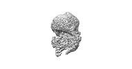
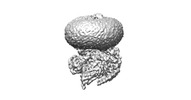
| Images |  emd_37390.png emd_37390.png | 34.6 KB | ||
| Filedesc metadata | emd-37390.cif.gz | 6.8 KB | ||
| Others | emd_37390_half_map_1.map.gzemd_37390_half_map_2.map.gz | 59.3 MB 59.3 MB | ||
| Archive directory |  http://ftp.pdbj.org/pub/emdb/structures/EMD-37390ftp://ftp.pdbj.org/pub/emdb/structures/EMD-37390 http://ftp.pdbj.org/pub/emdb/structures/EMD-37390ftp://ftp.pdbj.org/pub/emdb/structures/EMD-37390 | HTTPS FTP |
-Related structure data
| Related structure data | 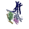 8wa3MC 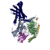 8wg7C 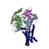 8wg8C M: atomic model generated by this map C: citing same article ( |
|---|---|
| Similar structure data |
-Links
| EMDB pages | EMDB (EBI/PDBe) / EMDataResource |
|---|---|
| Related items in Molecule of the Month |


-Map
| File | Download / File: emd_37390.map.gz / Format: CCP4 / Size: 64 MB / Type: IMAGE STORED AS FLOATING POINT NUMBER (4 BYTES) | ||||||||||||||||||||
|---|---|---|---|---|---|---|---|---|---|---|---|---|---|---|---|---|---|---|---|---|---|
| Voxel size | X=Y=Z: 1.071 Å | ||||||||||||||||||||
| Density |
| ||||||||||||||||||||
| Symmetry | Space group: 1 | ||||||||||||||||||||
| Details | EMDB XML:
|
-Supplemental data
-Half map: #2
| File | emd_37390_half_map_1.map | ||||||||||||
|---|---|---|---|---|---|---|---|---|---|---|---|---|---|
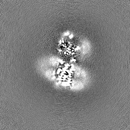
| Projections & Slices |
| ||||||||||||
| Density Histograms |
 Z
Z Y
Y X
X


-Half map: #1
| File | emd_37390_half_map_2.map | ||||||||||||
|---|---|---|---|---|---|---|---|---|---|---|---|---|---|
| Projections & Slices |
| ||||||||||||
| Density Histograms |


- Sample components
-Entire : Cryo-EM structure of the human glucose-dependent insulinotropic p...
| Entire | Name: Cryo-EM structure of the human glucose-dependent insulinotropic polypeptide receptor in complex with GIP and G protein |
|---|---|
| Components |
|
-Supramolecule #1: Cryo-EM structure of the human glucose-dependent insulinotropic p...
| Supramolecule | Name: Cryo-EM structure of the human glucose-dependent insulinotropic polypeptide receptor in complex with GIP and G protein type: complex / ID: 1 / Parent: 0 / Macromolecule list: all |
|---|---|
| Source (natural) | Organism: Homo sapiens (human) |
-Macromolecule #1: Gastric inhibitory polypeptide receptor,Fusion protein
| Macromolecule | Name: Gastric inhibitory polypeptide receptor,Fusion protein type: protein_or_peptide / ID: 1 / Number of copies: 1 / Enantiomer: LEVO |
|---|---|
| Source (natural) | Organism: Homo sapiens (human) |
| Molecular weight | Theoretical: 64.950695 KDa |
| Recombinant expression | Organism:   Spodoptera frugiperda (fall armyworm) Spodoptera frugiperda (fall armyworm) |
| Sequence | String: RAETGSKGQT AGELYQRWER YRRECQETLA AAEPPSGLAC NGSFDMYVCW DYAAPNATAR ASCPWYLPWH HHVAAGFVLR QCGSDGQWG LWRDHTQCEN PEKNEAFLDQ RLILERLQVM YTVGYSLSLA TLLLALLILS LFRRLHCTRN YIHINLFTSF M LRAAAILS ...String: RAETGSKGQT AGELYQRWER YRRECQETLA AAEPPSGLAC NGSFDMYVCW DYAAPNATAR ASCPWYLPWH HHVAAGFVLR QCGSDGQWG LWRDHTQCEN PEKNEAFLDQ RLILERLQVM YTVGYSLSLA TLLLALLILS LFRRLHCTRN YIHINLFTSF M LRAAAILS RDRLLPRPGP YLGDQALALW NQALAACRTA QIVTQYCVGA NYTWLLVEGV YLHSLLVLVG GSEEGHFRYY LL LGWGAPA LFVIPWVIVR YLYENTQCWE RNEVKAIWWI IRTPILMTIL INFLIFIRIL GILLSKLRTR QMRCRDYRLR LAR STLFLV PLLGVHEVVF APVTEEQARG ALRFAKLGFE IFLSSFQGFL VSVLYCFINK EVQSEIRRGW HHCRLRRSLG EEQR GSSGG GGSGGGGSSG VFTLEDFVGD WEQTAAYNLD QVLEQGGVSS LLQNLAVSVT PIQRIVRSGE NALKIDIHVI IPYEG LSAD QMAQIEEVFK VVYPVDDHHF KVILPYGTLV IDGVTPNMLN YFGRPYEGIA VFDGKKITVT GTLWNGNKII DERLIT PDG SMLFRVTINS UniProtKB: Gastric inhibitory polypeptide receptor |
-Macromolecule #2: Guanine nucleotide-binding protein G(s) subunit alpha isoforms short
| Macromolecule | Name: Guanine nucleotide-binding protein G(s) subunit alpha isoforms short type: protein_or_peptide / ID: 2 / Number of copies: 1 / Enantiomer: LEVO |
|---|---|
| Source (natural) | Organism: Bos taurus (cattle) |
| Molecular weight | Theoretical: 45.743441 KDa |
| Recombinant expression | Organism: Spodoptera frugiperda (fall armyworm) |
| Sequence | String: MGCLGNSKTE DQRNEEKAQR EANKKIEKQL QKDKQVYRAT HRLLLLGAGE SGKNTIVKQM RILHVNGFNG EGGEEDPQAA RSNSDGEKA TKVQDIKNNL KEAIETIVAA MSNLVPPVEL ANPENQFRVD YILSVMNVPD FDFPPEFYEH AKALWEDEGV R ACYERSNE ...String: MGCLGNSKTE DQRNEEKAQR EANKKIEKQL QKDKQVYRAT HRLLLLGAGE SGKNTIVKQM RILHVNGFNG EGGEEDPQAA RSNSDGEKA TKVQDIKNNL KEAIETIVAA MSNLVPPVEL ANPENQFRVD YILSVMNVPD FDFPPEFYEH AKALWEDEGV R ACYERSNE YQLIDCAQYF LDKIDVIKQD DYVPSDQDLL RCRVLTSGIF ETKFQVDKVN FHMFDVGAQR DERRKWIQCF ND VTAIIFV VASSSYNMVI REDNQTNRLQ AALKLFDSIW NNKWLRDTSV ILFLNKQDLL AEKVLAGKSK IEDYFPEFAR YTT PEDATP EPGEDPRVTR AKYFIRDEFL RISTASGDGR HYCYPHFTCS VDTENIRRVF NDCRDIIQRM HLRQYELL UniProtKB: Guanine nucleotide-binding protein G(s) subunit alpha isoforms short |
-Macromolecule #3: Guanine nucleotide-binding protein G(I)/G(S)/G(T) subunit beta-1,...
| Macromolecule | Name: Guanine nucleotide-binding protein G(I)/G(S)/G(T) subunit beta-1,O-antigen polymerase type: protein_or_peptide / ID: 3 / Number of copies: 1 / Enantiomer: LEVO |
|---|---|
| Source (natural) | Organism: Leptolinea tardivitalis (bacteria) |
| Molecular weight | Theoretical: 40.226992 KDa |
| Recombinant expression | Organism: Spodoptera frugiperda (fall armyworm) |
| Sequence | String: MGSLLQSELD QLRQEAEQLK NQIRDARKAC ADATLSQITN NIDPVGRIQM RTRRTLRGHL AKIYAMHWGT DSRLLVSASQ DGKLIIWDS YTTNKVHAIP LRSSWVMTCA YAPSGNYVAC GGLDNICSIY NLKTREGNVR VSRELAGHTG YLSCCRFLDD N QIVTSSGD ...String: MGSLLQSELD QLRQEAEQLK NQIRDARKAC ADATLSQITN NIDPVGRIQM RTRRTLRGHL AKIYAMHWGT DSRLLVSASQ DGKLIIWDS YTTNKVHAIP LRSSWVMTCA YAPSGNYVAC GGLDNICSIY NLKTREGNVR VSRELAGHTG YLSCCRFLDD N QIVTSSGD TTCALWDIET GQQTTTFTGH TGDVMSLSLA PDTRLFVSGA CDASAKLWDV REGMCRQTFT GHESDINAIC FF PNGNAFA TGSDDATCRL FDLRADQELM TYSHDNIICG ITSVSFSKSG RLLLAGYDDF NCNVWDALKA DRAGVLAGHD NRV SCLGVT DDGMAVATGS WDSFLKIWNG SSGGGGSGGG GSSGVSGWRL FKKIS UniProtKB: Guanine nucleotide-binding protein G(I)/G(S)/G(T) subunit beta-1, O-antigen polymerase |
-Macromolecule #4: Guanine nucleotide-binding protein G(I)/G(S)/G(O) subunit gamma-2
| Macromolecule | Name: Guanine nucleotide-binding protein G(I)/G(S)/G(O) subunit gamma-2 type: protein_or_peptide / ID: 4 / Number of copies: 1 / Enantiomer: LEVO |
|---|---|
| Source (natural) | Organism: Bos taurus (cattle) |
| Molecular weight | Theoretical: 7.861143 KDa |
| Recombinant expression | Organism: Spodoptera frugiperda (fall armyworm) |
| Sequence | String: MASNNTASIA QARKLVEQLK MEANIDRIKV SKAAADLMAY CEAHAKEDPL LTPVPASENP FREKKFFCAI L UniProtKB: Guanine nucleotide-binding protein G(I)/G(S)/G(O) subunit gamma-2 |
-Macromolecule #5: Nanobody-35
| Macromolecule | Name: Nanobody-35 / type: protein_or_peptide / ID: 5 / Number of copies: 1 / Enantiomer: LEVO |
|---|---|
| Source (natural) | Organism: synthetic construct (others) |
| Molecular weight | Theoretical: 15.343019 KDa |
| Recombinant expression | Organism: Escherichia coli (E. coli) |
| Sequence | String: MAQVQLQESG GGLVQPGGSL RLSCAASGFT FSNYKMNWVR QAPGKGLEWV SDISQSGASI SYTGSVKGRF TISRDNAKNT LYLQMNSLK PEDTAVYYCA RCPAPFTRDC FDVTSTTYAY RGQGTQVTVS SHHHHHHEPE A |
-Experimental details
-Structure determination
| Method | cryo EM |
|---|---|
 Processing Processing | single particle reconstruction |
| Aggregation state | particle |
-Sample preparation
| Buffer | pH: 7.4 |
|---|---|
| Vitrification | Cryogen name: ETHANE |
| Details | Cryo-EM structure of peptide free and Gs-coupled GIPR |
- Electron microscopy
| Microscope | FEI TITAN KRIOS |
|---|---|
| Electron beam | Acceleration voltage: 300 kV / Electron source: OTHER |
| Electron optics | Illumination mode: OTHER / Imaging mode: BRIGHT FIELDBright-field microscopy / Nominal defocus max: 2.2 µm / Nominal defocus min: 1.2 µm |
| Image recording | Film or detector model: GATAN K3 (6k x 4k) / Average electron dose: 80.0 e/Å2 |
| Experimental equipment |  Model: Titan Krios / Image courtesy: FEI Company |
-Image processing
| Startup model | Type of model: PDB ENTRY PDB model - PDB ID: Details: and 2QKH |
|---|---|
| Initial angle assignment | Type: OTHER |
| Final angle assignment | Type: OTHER |
| Final reconstruction | Resolution.type: BY AUTHOR / Resolution: 2.86 Å / Resolution method: OTHER / Number images used: 807839 |