Francesca Vallese / Kookjoo Kim / Laura Y Yen / Jake D Johnston / Alex J Noble / Tito Calì / Oliver Biggs Clarke /
PubMed Abstract
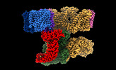
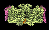
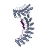
The stability and shape of the erythrocyte membrane is provided by the ankyrin-1 complex, but how it tethers the spectrin-actin cytoskeleton to the lipid bilayer and the nature of its association ...The stability and shape of the erythrocyte membrane is provided by the ankyrin-1 complex, but how it tethers the spectrin-actin cytoskeleton to the lipid bilayer and the nature of its association with the band 3 anion exchanger and the Rhesus glycoproteins remains unknown. Here we present structures of ankyrin-1 complexes purified from human erythrocytes. We reveal the architecture of a core complex of ankyrin-1, the Rhesus proteins RhAG and RhCE, the band 3 anion exchanger, protein 4.2, glycophorin A and glycophorin B. The distinct T-shaped conformation of membrane-bound ankyrin-1 facilitates recognition of RhCE and, unexpectedly, the water channel aquaporin-1. Together, our results uncover the molecular details of ankyrin-1 association with the erythrocyte membrane, and illustrate the mechanism of ankyrin-mediated membrane protein clustering.
EMDB-26886, PDB-7uze: Erythrocyte ankyrin-1 complex class 2 local refinement of AQP1 (C4 symmetry applied) Method: EM (single particle) / Resolution: 2.4 Å
EMDB-26916, PDB-7uzq: Local refinement of RhAG-RhCE-ANK1(AR1-5), from consensus refinement of all classes Method: EM (single particle) / Resolution: 2.17 Å
EMDB-26917, PDB-7uzs: Protein 4.2 (local refinement from consensus reconstruction of ankyrin complex classes) Method: EM (single particle) / Resolution: 2.2 Å
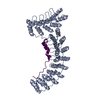
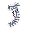
EMDB-26918, PDB-7uzu: Ankyrin-1 (N-terminal region of membrane binding domain, local refinement from consensus reconstruction; bound to N-terminal peptide from band 3) Method: EM (single particle) / Resolution: 2.3 Å
EMDB-26919, PDB-7uzv: Cytoplasmic domains of Band 3-I (local refinement from consensus reconstruction of ankyrin complexes) Method: EM (single particle) / Resolution: 2.5 Å
EMDB-26940, PDB-7v07: Band 3-I-TM local refinement from erythrocyte ankyrin-1 complex consensus reconstruction Method: EM (single particle) / Resolution: 2.8 Å
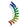
EMDB-26944, PDB-7v0m: Local refinement of ankyrin-1 (N-terminal half), class 1 of erythrocyte ankyrin-1 complex Method: EM (single particle) / Resolution: 2.7 Å
EMDB-26950, PDB-7v0t: Local refinement of Band 3-I cytoplasmic domains, class 1 of erythrocyte ankyrin-1 complex Method: EM (single particle) / Resolution: 2.7 Å
EMDB-26951, PDB-7v0u: Local refinement of Band 3-II cytoplasmic domains, class 1 of erythrocyte ankyrin-1 complex Method: EM (single particle) / Resolution: 3.0 Å
EMDB-26952, PDB-7v0x: Local refinement of ankyrin-1 (C-terminal half), class 1 of erythrocyte ankyrin-1 complex Method: EM (single particle) / Resolution: 3.0 Å
EMDB-26953, PDB-7v0y: Local refinement of Band 3-III cytoplasmic domains, class 1 of erythrocyte ankyrin-1 complex Method: EM (single particle) / Resolution: 3.0 Å
EMDB-26954, PDB-7v19: Local refinement of Band 3-II transmembrane domains, class 1 of erythrocyte ankyrin-1 complex Method: EM (single particle) / Resolution: 3.3 Å
EMDB-26955, PDB-8crq: Local refinement of Band 3-I transmembrane domains, class 1 of erythrocyte ankyrin-1 complex Method: EM (single particle) / Resolution: 3.2 Å
EMDB-26956, PDB-8crr: Local refinement of Band 3-III transmembrane domains, class 1 of erythrocyte ankyrin-1 complex Method: EM (single particle) / Resolution: 3.0 Å
EMDB-26958, PDB-8crt: Local refinement of Rh trimer, glycophorin B and Band3-III transmembrane region, class 1a of erythrocyte ankyrin-1 complex Method: EM (single particle) / Resolution: 3.0 Å
EMDB-26972, PDB-8csv: Local refinement of Anykyrin-1 (N-terminal half of membrane binding domain) in Class 2 of erythrocyte ankyrin-1 complex Method: EM (single particle) / Resolution: 2.7 Å
EMDB-26975, PDB-8csy: Local refinement of cytoplasmic domains of band3-I in class 2 of erythrocyte ankyrin-1 complex Method: EM (single particle) / Resolution: 2.7 Å
EMDB-26978, PDB-8ct2: Local refinement of AQP1 tetramer (C1; refinement mask included D1 of protein 4.2 and Ankyrin-1 AR1-5) in Class 2 of erythrocyte ankyrin-1 complex Method: EM (single particle) / Resolution: 3.1 Å
EMDB-26979, PDB-8ct3: Local refinement of band3-I transmembrane region from class 2 of erythrocyte ankyrin-1 complex Method: EM (single particle) / Resolution: 3.3 Å
TRANSPORT PROTEIN / Membrane Protein / Anion Exchange / Erythrocyte / Glycoprotein / STRUCTURAL PROTEIN / Ankyrin / TRANSPORT PROTEIN/STRUCTURAL PROTEIN / TRANSPORT PROTEIN-STRUCTURAL PROTEIN complex / ankyrin complex
+
About Yorodumi Papers
-
News
-
Feb 9, 2022. New format data for meta-information of EMDB entries
New format data for meta-information of EMDB entries
Version 3 of the EMDB header file is now the official format.
The previous official version 1.9 will be removed from the archive.
In the structure databanks used in Yorodumi, some data are registered as the other names, "COVID-19 virus" and "2019-nCoV". Here are the details of the virus and the list of structure data.
Jan 31, 2019. EMDB accession codes are about to change! (news from PDBe EMDB page)
EMDB accession codes are about to change! (news from PDBe EMDB page)
The allocation of 4 digits for EMDB accession codes will soon come to an end. Whilst these codes will remain in use, new EMDB accession codes will include an additional digit and will expand incrementally as the available range of codes is exhausted. The current 4-digit format prefixed with “EMD-” (i.e. EMD-XXXX) will advance to a 5-digit format (i.e. EMD-XXXXX), and so on. It is currently estimated that the 4-digit codes will be depleted around Spring 2019, at which point the 5-digit format will come into force.
The EM Navigator/Yorodumi systems omit the EMD- prefix.
Related info.:Q: What is EMD? / ID/Accession-code notation in Yorodumi/EM Navigator
 Movie
Movie Controller
 Structure viewers
 About Yorodumi Papers


 Authors

 External links


 Keywords
 TRANSPORT PROTEIN /