 ムービー
ムービー コントローラー
 構造ビューア
 EMN検索について


-検索条件
-検索結果
検索 (著者・登録者: bukau & b)の結果60件中、1から50件目までを表示しています
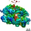
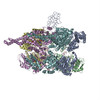
EMDB-15311: 
Dedicated chaperone at the ribosome safeguards the proteostasis network during eEF1A biogenesis
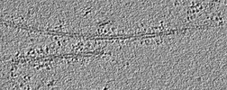
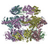
EMDB-13576:
Alpha-synuclein amyloid fibres incubated with DNAJB1, Hsc70, Hsp110 and ATP
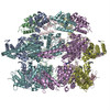
EMDB-13577:
Alpha-synuclein amyloid fibres incubated with DNAJB1, Hsc70, and ATP

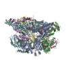
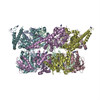
EMDB-14156:
Cryo-EM reconstruction of the Bacillus subtilis MutS2-collided disome complex (MutS2 conf.1; Leading 70S)

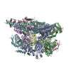
EMDB-14157:
Composite reconstruction of the Bacillus subtilis collided disome (Leading 70S)

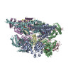
EMDB-14158:
Composite reconstruction of the Bacillus subtilis collided disome (Collided 70S)

EMDB-14159:
Cryo-EM reconstruction of the Bacillus subtilis MutS2-collided disome complex (MutS2 conf.2; Leading 70S)

EMDB-14160:
Cryo-EM reconstruction of the Bacillus subtilis MutS2-collided disome complex (Leading 70S)

EMDB-14161:
Cryo-EM reconstruction of the Bacillus subtilis MutS2-collided disome complex (Collided 70S)

EMDB-14162:
Cryo-EM reconstruction of the Bacillus subtilis collided disome (Leading 70S)

EMDB-14163:
Cryo-EM reconstruction of the Bacillus subtilis collided disome (Leading 30S)

EMDB-14164:
Cryo-EM reconstruction of the Bacillus subtilis collided disome (Collided 70S)

EMDB-14165:
Cryo-EM reconstruction of the Bacillus subtilis collided disome (Collided 30S)

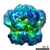
EMDB-14166:
Cryo-EM reconstruction of Bacillus subtilis obstructed 50S subunit co-purified with MutS2

PDB-7qv1:
Bacillus subtilis collided disome (Leading 70S)

PDB-7qv2:
Bacillus subtilis collided disome (Collided 70S)

PDB-7qv3:
Bacillus subtilis MutS2-collided disome complex (MutS2 conf.2; Leading 70S)
EMDB-4621:
ClpB (DWB and K476C mutant) bound to casein in presence of ATPgammaS - state KC-3
EMDB-4622:
E. coli ClpB (DWB and K476C mutant) bound to casein - middle domain conformation 1
EMDB-4623:
E. coli ClpB (DWB and K476C mutant) bound to casein - middle domain conformation 2
EMDB-4624:
ClpB (DWB and K476C mutant) bound to casein in presence of ATPgammaS - state KC-1
EMDB-4625:
E. coli ClpB (DWB and K476C mutant) bound to casein - state KC-2
EMDB-4626:
ClpB (DWB and K476C mutant) bound to casein in presence of ATPgammaS - state KC-2A
EMDB-4627:
ClpB (DWB and K476C mutant) bound to casein in presence of ATPgammaS - state KC-2B
EMDB-4940:
ClpB (DWB mutant) bound to casein in presence of ATPgammaS - state WT-1

EMDB-4941:
ClpB (DWB mutant) bound to casein in presence of ATPgammaS - state WT-2A
EMDB-4942:
ClpB (DWB mutant) bound to casein in presence of ATPgammaS - state WT-2B
PDB-6qs4:
Two-Step Activation Mechanism of the ClpB Disaggregase for Sequential Substrate Threading by the Main ATPase Motor.
PDB-6qs6:
ClpB (DWB and K476C mutant) bound to casein in presence of ATPgammaS - state KC-1
PDB-6qs7:
ClpB (DWB and K476C mutant) bound to casein in presence of ATPgammaS - state KC-2A
PDB-6qs8:
ClpB (DWB and K476C mutant) bound to casein in presence of ATPgammaS - state KC-2B
PDB-6rn2:
ClpB (DWB mutant) bound to casein in presence of ATPgammaS - state WT-1
PDB-6rn3:
ClpB (DWB mutant) bound to casein in presence of ATPgammaS - state WT-2A
PDB-6rn4:
ClpB (DWB mutant) bound to casein in presence of ATPgammaS - state WT-2B

EMDB-3894:
S.aureus ClpC resting state, C2 symmetrised

EMDB-3895:
S.aureus ClpC resting state, asymmetric map
EMDB-3897:
Structure of S.aureus ClpC in complex with MecA

PDB-6em8:
S.aureus ClpC resting state, C2 symmetrised

PDB-6em9:
S.aureus ClpC resting state, asymmetric map
PDB-6emw:
Structure of S.aureus ClpC in complex with MecA
EMDB-3776:
Cryo EM structure of the bacterial disaggregase ClpB (BAP form, DWB mutant), in the ATPgammaS state, bound to the model substrate casein
EMDB-3777:
Cryo EM structure of the bacterial disaggregase ClpB (BAP form, DWB mutant), in the ATPgammaS state
PDB-5ofo:
Cryo EM structure of the E. coli disaggregase ClpB (BAP form, DWB mutant), in the ATPgammaS state, bound to the model substrate casein
PDB-5og1:
Cryo EM structure of the E. coli disaggregase ClpB (BAP form, DWB mutant), in the ATPgammaS state

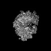
EMDB-3094:
Electron negative stain tomography of alpha-synuclein amyloid fibrils

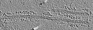
EMDB-3095:
Electron negative stain tomography of alpha-synuclein amyloid fibrils treated with the Hsc70/DNAJB1/Apg2 chaperone system
PDB-4d2q:
Negative-stain electron microscopy of E. coli ClpB mutant E432A (BAP form bound to ClpP)
PDB-4d2u:
Negative-stain electron microscopy of E. coli ClpB (BAP form bound to ClpP)
PDB-4d2x:
Negative-stain electron microscopy of E. coli ClpB of Y503D hyperactive mutant (BAP form bound to ClpP)
EMDB-2555:
Negative-stain electron microscopy of E. coli ClpB mutant E432A (BAP form bound to ClpP)
ページ:
 wwPDBはEMDBデータモデルのバージョン3へ移行します
