 Movie
Movie Controller
 Structure viewers
 About EMN search


-Search query
-Search result
Showing 1 - 50 of 1,883 items for (author: j. & lin)

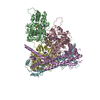
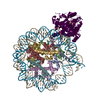
PDB-8qv0: 
Structure of the native microtubule lattice nucleated from the yeast spindle pole body
Method: subtomogram averaging / : Dendooven T, Yatskevich S, Burt A, Bellini D, Kilmartin J, Barford D

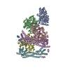
PDB-8qv2:
Structure of the native y-Tubulin Ring Complex (yTuRC) capping microtubule minus ends at the spindle pole body
Method: subtomogram averaging / : Dendooven T, Yatskevich S, Burt A, Bellini D, Kilmartin J, Barford D

PDB-8qv3:
Structure of the y-Tubulin Small Complex (yTuSC) as part of the native y-Tubulin Ring Complex (yTuRC) capping microtubule minus ends at the spindle pole body
Method: subtomogram averaging / : Dendooven T, Yatskevich S, Burt A, Bellini D, Kilmartin J, Barford D

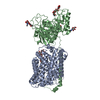
PDB-8kdb:
Cryo-EM structure of the human parainfluenza virus hPIV3 L-P polymerase in dimeric form
Method: single particle / : Xie J, Wang L, Zhai G, Wu D, Lin Z, Wang M, Yan X, Gao L, Huang X, Fearns R, Chen S

PDB-8kdc:
Cryo-EM structure of the human parainfluenza virus hPIV3 L-P polymerase in monomeric form
Method: single particle / : Xie J, Wang L, Zhai G, Wu D, Lin Z, Wang M, Yan X, Gao L, Huang X, Fearns R, Chen S

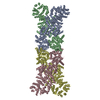
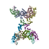
PDB-8rhz:
Structure of CUL9-RBX1 ubiquitin E3 ligase complex in unneddylated conformation - symmetry expanded unneddylated dimer
Method: single particle / : Hopf LVM, Horn-Ghetko D, Prabu JR, Schulman BA

PDB-8u6y:
Preholo-Proteasome from Beta 3 D205 deletion
Method: single particle / : Walsh Jr RM, Rawson S, Velez B, Blickling M, Razi A, Hanna J
PDB-8u7u:
Proteasome 20S Core Particle from Beta 3 D205 deletion
Method: single particle / : Walsh Jr RM, Rawson S, Velez B, Blickling M, Razi A, Hanna J

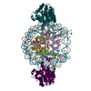
PDB-9asb:
Structure of human calcium-sensing receptor in complex with chimeric Gq (miniGisq) protein in nanodiscs
Method: single particle / : Zuo H, Park J, Frangaj A, Ye J, Lu G, Manning JJ, Asher WB, Lu Z, Hu G, Wang L, Mendez J, Eng E, Zhang Z, Lin X, Grasucci R, Hendrickson WA, Clarke OB, Javitch JA, Conigrave AD, Fan QR

PDB-9avg:
Structure of human calcium-sensing receptor in complex with chimeric Gs (miniGis) protein in nanodiscs
Method: single particle / : Zuo H, Park J, Frangaj A, Ye J, Lu G, Manning JJ, Asher WB, Lu Z, Hu G, Wang L, Mendez J, Eng E, Zhang Z, Lin X, Grasucci R, Hendrickson WA, Clarke OB, Javitch JA, Conigrave AD, Fan QR

PDB-9avl:
Structure of human calcium-sensing receptor in complex with Gi3 protein in nanodiscs
Method: single particle / : Zuo H, Park J, Frangaj A, Ye J, Lu G, Manning JJ, Asher WB, Lu Z, Hu G, Wang L, Mendez J, Eng E, Zhang Z, Lin X, Grasucci R, Hendrickson WA, Clarke OB, Javitch JA, Conigrave AD, Fan QR
PDB-9axf:
Structure of human calcium-sensing receptor in complex with chimeric Gq (miniGisq) protein in detergent
Method: single particle / : Zuo H, Park J, Frangaj A, Ye J, Lu G, Manning JJ, Asher WB, Lu Z, Hu G, Wang L, Mendez J, Eng E, Zhang Z, Lin X, Grasucci R, Hendrickson WA, Clarke OB, Javitch JA, Conigrave AD, Fan QR
PDB-9ayf:
Structure of human calcium-sensing receptor in complex with Gi1 (miniGi1) protein in detergent
Method: single particle / : Zuo H, Park J, Frangaj A, Ye J, Lu G, Manning JJ, Asher WB, Lu Z, Hu G, Wang L, Mendez J, Eng E, Zhang Z, Lin X, Grasucci R, Hendrickson WA, Clarke OB, Javitch JA, Conigrave AD, Fan QR

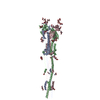
PDB-8rty:
Structure of the F-actin barbed end bound by Cdc12 and profilin (ring complex) at a resolution of 6.3 Angstrom
Method: single particle / : Oosterheert W, Boiero Sanders M, Funk J, Prumbaum D, Raunser S, Bieling P
PDB-8ru0:
Structure of the undecorated barbed end of F-actin.
Method: single particle / : Oosterheert W, Boiero Sanders M, Funk J, Prumbaum D, Raunser S, Bieling P
PDB-8ru2:
Structure of the F-actin barbed end bound by formin mDia1
Method: single particle / : Oosterheert W, Boiero Sanders M, Funk J, Prumbaum D, Raunser S, Bieling P
PDB-8rv2:
Structure of the formin INF2 bound to the barbed end of F-actin.
Method: single particle / : Oosterheert W, Boiero Sanders M, Funk J, Prumbaum D, Raunser S, Bieling P
PDB-8qey:
Structure of human Asc1/CD98hc heteromeric amino acid transporter
Method: single particle / : Martinez-Molledo M, Rullo-Tubau J, Errasti-Murugarren E, Palacin M, Llorca O
PDB-8wy9:
Cryo-EM structure of DSR2 apo (partial) complex
Method: single particle / : Zhang JT, Jia N, Liu XY
PDB-8wyb:
Cryo-EM structure of DSR2 (H171A)-tube-NAD+ complex
Method: single particle / : Zhang JT, Jia N, Liu XY
PDB-8wyc:
Cryo-EM structure of DSR2 (H171A)-tube-NAD+ (partial) complex
Method: single particle / : Zhang JT, Jia N, Liu XY
PDB-8wyd:
Cryo-EM structure of DSR2-DSAD1 complex
Method: single particle / : Zhang JT, Jia N, Liu XY
PDB-8wye:
Cryo-EM structure of DSR2-DSAD1 (partial) complex
Method: single particle / : Zhang JT, Jia N, Liu XY
PDB-8wyf:
Cryo-EM structure of DSR2-DSAD1-NAD+ (partial) complex
Method: single particle / : Zhang JT, Jia N, Liu XY

PDB-8wa2:
cryo-EM structure of native mastigonemes isolated from Chlamydomonas reinhardtii at 3.0 angstrom resolution
Method: single particle / : Huang J, Tao H, Chen J, Pan J, Yan C, Yan N
PDB-8rtt:
Structure of the formin Cdc12 bound to the barbed end of phalloidin-stabilized F-actin.
Method: single particle / : Oosterheert W, Boiero Sanders M, Funk J, Prumbaum D, Raunser S, Bieling P
PDB-8v4y:
Cryo-EM structure of singly-bound SNF2h-nucleosome complex with SNF2h at inactive SHL2 (conformation 1)
Method: single particle / : Chio US, Palovcak E, Armache JP, Narlikar GJ, Cheng Y
PDB-8v6v:
Cryo-EM structure of doubly-bound SNF2h-nucleosome complex
Method: single particle / : Chio US, Palovcak E, Armache JP, Narlikar GJ, Cheng Y

PDB-8v7l:
Cryo-EM structure of singly-bound SNF2h-nucleosome complex with SNF2h at inactive SHL2 (conformation 2)
Method: single particle / : Chio US, Palovcak E, Armache JP, Narlikar GJ, Cheng Y
PDB-7xog:
Cryo-EM structure of S glycoprotein encoded by the Covid-19 mRNA vaccine candidate RQ3013 (Postfusion state)
Method: single particle / : Wu Z, Yu Z, Tan S, Lu J, Lu G, Lin J
PDB-8jay:
CrtSPARTA Octamer bound with guide-target
Method: single particle / : Guo LJ, Huang PP, Li ZX, Xiao YB, Chen MR
PDB-8tum:
Type IV pilus from Pseudomonas PAO1 strain
Method: single particle / : Thongchol J, Zhang J, Zeng L
PDB-8tuw:
Type IV pilus from Pseudomonas PAO1 strain with PP7 Maturation protein
Method: single particle / : Thongchol J, Zhang J, Zeng L

PDB-8tux:
Capsid of mature PP7 virion with 3'end region of PP7 genomic RNA
Method: single particle / : Thongchol J, Zhang J, Zeng L
PDB-8oqi:
Cryo-EM structure of the wild-type alpha-synuclein fibril.
Method: helical / : Pesch V, Reithofer S, Ma L, Flores-Fernandez JM, Oezduezenciler P, Busch Y, Lien Y, Rudtke O, Frieg B, Schroeder GF, Wille H, Tamgueney G

PDB-8tz1:
Cryo-EM structure of bovine concentrative nucleoside transporter 3 in complex with Ribavirin
Method: single particle / : Wright NJ, Lee SY
PDB-8tz2:
Cryo-EM structure of bovine concentrative nucleoside transporter 3 in MSP2N2 nanodiscs, apo state
Method: single particle / : Wright NJ, Lee SY
PDB-8tz3:
Cryo-EM structure of bovine concentrative nucleoside transporter 3 in complex with GS-441524, consensus reconstruction
Method: single particle / : Wright NJ, Lee SY
PDB-8tz4:
Cryo-EM structure of bovine concentrative nucleoside transporter 3 in complex with GS-441524, subset reconstruction
Method: single particle / : Wright NJ, Lee SY
PDB-8tz5:
Cryo-EM structure of bovine concentrative nucleoside transporter 3 in complex with N-hydroxycytidine
Method: single particle / : Wright NJ, Lee SY
PDB-8tz6:
Cryo-EM structure of bovine concentrative nucleoside transporter 3 in complex with PSI-6206
Method: single particle / : Wright NJ, Lee SY
PDB-8tz7:
Cryo-EM structure of bovine concentrative nucleoside transporter 3 in complex with Molnupiravir, condition 1, INT1-INT1-INT1 conformation
Method: single particle / : Wright NJ, Lee SY
PDB-8tz8:
Cryo-EM structure of bovine concentrative nucleoside transporter 3 in complex with Molnupiravir, condition 1, INT1-INT1-INT3 conformation
Method: single particle / : Wright NJ, Lee SY
PDB-8tz9:
Cryo-EM structure of bovine concentrative nucleoside transporter 3 in complex with Molnupiravir, condition 2, INT2-INT2-INT2 conformation
Method: single particle / : Wright NJ, Lee SY
PDB-8tza:
Cryo-EM structure of bovine concentrative nucleoside transporter 3 in complex with Molnupiravir, condition 2, INT2-INT1-INT1 conformation
Method: single particle / : Wright NJ, Lee SY
PDB-8tzd:
Cryo-EM structure of bovine concentrative nucleoside transporter 3 in complex with Molnupiravir, condition 1, INT1-INT1-OFS conformation (3DVA analysis)
Method: single particle / : Wright NJ, Lee SY
PDB-8w5k:
Cryo-EM structure of the yeast TOM core complex crosslinked by BS3 (from TOM-TIM23 complex)
Method: single particle / : Wang Q, Guan ZY, Zhuang JJ, Huang R, Yin P
PDB-8j84:
Short ago complexed with TIR-APAZ
Method: single particle / : Guo LJ, Huang PP, Li ZX, Xiao YB, Chen MR
Pages:
 wwPDB to switch to version 3 of the EMDB data model
