Movie
Movie Controller

[English] 日本語
 Yorodumi
- PDB-9avl: Structure of human calcium-sensing receptor in complex with Gi3 p... -
+ Open data

- Basic information
| Entry | Database: PDB / ID: 9avl | ||||||
|---|---|---|---|---|---|---|---|
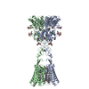
| Title | Structure of human calcium-sensing receptor in complex with Gi3 protein in nanodiscs | ||||||
 Components Components |
| ||||||
 Keywords Keywords |  MEMBRANE PROTEIN / Calcium-sensing receptor / G-protein-coupled receptor / G protein / signal transduction MEMBRANE PROTEIN / Calcium-sensing receptor / G-protein-coupled receptor / G protein / signal transduction | ||||||
| Function / homology |  Function and homology information Function and homology informationbile acid secretion / chemosensory behavior / cellular response to peptide / response to fibroblast growth factor / cellular response to vitamin D / phosphatidylinositol phospholipase C activity / negative regulation of adenylate cyclase activity / Class C/3 (Metabotropic glutamate/pheromone receptors) / regulation of potassium ion transmembrane transport / calcium ion import ...bile acid secretion / chemosensory behavior / cellular response to peptide / response to fibroblast growth factor / cellular response to vitamin D / phosphatidylinositol phospholipase C activity / negative regulation of adenylate cyclase activity / Class C/3 (Metabotropic glutamate/pheromone receptors) / regulation of potassium ion transmembrane transport / calcium ion import / GTP metabolic process / positive regulation of positive chemotaxis / fat pad development / dopamine receptor signaling pathway / amino acid binding / cellular response to hepatocyte growth factor stimulus / branching morphogenesis of an epithelial tube / positive regulation of calcium ion import / regulation of calcium ion transport / positive regulation of macroautophagy / cellular response to low-density lipoprotein particle stimulus / Adenylate cyclase inhibitory pathway / detection of calcium ion / anatomical structure morphogenesis / axon terminus / positive regulation of vasoconstriction / JNK cascade / chloride transmembrane transport / adenylate cyclase-inhibiting G protein-coupled receptor signaling pathway / ossification / G protein-coupled receptor binding / G protein-coupled receptor activity / response to ischemia / cellular response to glucose stimulus / G-protein beta/gamma-subunit complex binding / G beta:gamma signalling through PLC beta / Presynaptic function of Kainate receptors / Thromboxane signalling through TP receptor / adenylate cyclase-modulating G protein-coupled receptor signaling pathway / G-protein activation / Activation of G protein gated Potassium channels / Inhibition of voltage gated Ca2+ channels via Gbeta/gamma subunits / Prostacyclin signalling through prostacyclin receptor / Glucagon signaling in metabolic regulation / G beta:gamma signalling through CDC42 / intracellular calcium ion homeostasis / ADP signalling through P2Y purinoceptor 12 / positive regulation of insulin secretion / G beta:gamma signalling through BTK / Adrenaline,noradrenaline inhibits insulin secretion / Glucagon-type ligand receptors / Vasopressin regulates renal water homeostasis via Aquaporins / G alpha (z) signalling events / cellular response to catecholamine stimulus / Glucagon-like Peptide-1 (GLP1) regulates insulin secretion / ADORA2B mediated anti-inflammatory cytokines production / vasodilation / adenylate cyclase-activating dopamine receptor signaling pathway / ADP signalling through P2Y purinoceptor 1 / G beta:gamma signalling through PI3Kgamma / cellular response to prostaglandin E stimulus / Cooperation of PDCL (PhLP1) and TRiC/CCT in G-protein beta folding / GPER1 signaling / GDP binding / G-protein beta-subunit binding / heterotrimeric G-protein complex / G alpha (12/13) signalling events / signaling receptor complex adaptor activity / Thrombin signalling through proteinase activated receptors (PARs) / integrin binding / GTPase binding / Ca2+ pathway / phospholipase C-activating G protein-coupled receptor signaling pathway / midbody / G alpha (i) signalling events / fibroblast proliferation / cellular response to hypoxia / G alpha (s) signalling events / G alpha (q) signalling events / basolateral plasma membrane / vesicle / transmembrane transporter binding / Extra-nuclear estrogen signaling / positive regulation of ERK1 and ERK2 cascade / cell cycle / G protein-coupled receptor signaling pathway / apical plasma membrane / cell division / lysosomal membrane / focal adhesion / GTPase activity / centrosome / neuronal cell body / calcium ion binding / positive regulation of cell population proliferation / protein-containing complex binding / GTP binding / positive regulation of gene expression / nucleolus / protein kinase bindingSimilarity search - Function | ||||||
| Biological species |  Homo sapiens (human) Homo sapiens (human) | ||||||
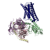
| Method | ELECTRON MICROSCOPY / single particle reconstruction / cryo EM / Resolution: 3.8 Å | ||||||
 Authors Authors | Zuo, H. / Park, J. / Frangaj, A. / Ye, J. / Lu, G. / Manning, J.J. / Asher, W.B. / Lu, Z. / Hu, G. / Wang, L. ...Zuo, H. / Park, J. / Frangaj, A. / Ye, J. / Lu, G. / Manning, J.J. / Asher, W.B. / Lu, Z. / Hu, G. / Wang, L. / Mendez, J. / Eng, E. / Zhang, Z. / Lin, X. / Grasucci, R. / Hendrickson, W.A. / Clarke, O.B. / Javitch, J.A. / Conigrave, A.D. / Fan, Q.R. | ||||||
| Funding support |  United States, 1items United States, 1items
| ||||||
 Citation Citation | Journal: Nature / Year: 2024 Title: Promiscuous G-protein activation by the calcium-sensing receptor Authors: Zuo, H. / Park, J. / Frangaj, A. / Ye, J. / Lu, G. / Manning, J.J. / Asher, W.B. / Lu, Z. / Hu, G. / Wang, L. / Mendez, J. / Eng, E. / Zhang, Z. / Lin, X. / Grasucci, R. / Hendrickson, W.A. ...Authors: Zuo, H. / Park, J. / Frangaj, A. / Ye, J. / Lu, G. / Manning, J.J. / Asher, W.B. / Lu, Z. / Hu, G. / Wang, L. / Mendez, J. / Eng, E. / Zhang, Z. / Lin, X. / Grasucci, R. / Hendrickson, W.A. / Clarke, O.B. / Javitch, J.A. / Conigrave, A.D. / Fan, Q.R. | ||||||
| History |
|
- Structure visualization
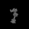
| Structure viewer | Molecule: MolmilJmol/JSmol |
|---|
- Downloads & links
-Download
| PDBx/mmCIF format | 9avl.cif.gz | 423.9 KB | Display | PDBx/mmCIF format |
|---|---|---|---|---|
| PDB format | pdb9avl.ent.gz | Display | PDB format | |
| PDBx/mmJSON format | 9avl.json.gz | Tree view | PDBx/mmJSON format | |
| Others |  Other downloads Other downloads |
-Validation report
| Arichive directory | https://data.pdbj.org/pub/pdb/validation_reports/av/9avlftp://data.pdbj.org/pub/pdb/validation_reports/av/9avl | HTTPS FTP |
|---|
-Related structure data
| Related structure data |  43908MC  9asbC  9avgC 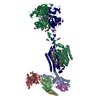 9axfC 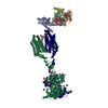 9ayfC C: citing same article ( M: map data used to model this data |
|---|---|
| Similar structure data |


-Links
 PDBj
PDBj


- Assembly
| Deposited unit | 
|
|---|---|
| 1 |
|
-Components
-Protein , 1 types, 2 molecules QR
| #1: Protein | Mass: 102864.617 Da / Num. of mol.: 2 Source method: isolated from a genetically manipulated source Details: The CaSR construct consists of residues 1-903 and a Flag tag inserted after the signal peptide. Source: (gene. exp.) Homo sapiens (human) / Gene: CASR, GPRC2A, PCAR1 / Cell line (production host): HEK293 GnTI- / Production host: Homo sapiens (human) / References: UniProt: P41180 |
|---|
-Guanine nucleotide-binding protein ... , 3 types, 3 molecules ABG
| #2: Protein | Mass: 40584.156 Da / Num. of mol.: 1 / Mutation: S47N, G203A, E245A, A326S Source method: isolated from a genetically manipulated source Details: G(i3)alpha contains four dominant negative mutations, S47N, G203A, E245A, and A326S Source: (gene. exp.) Homo sapiens (human) / Gene: GNAI3 / Cell line (production host): HEK293 GnTI- / Production host: Homo sapiens (human) / References: UniProt: P08754 |
|---|---|
| #3: Protein | Mass: 38367.969 Da / Num. of mol.: 1 Source method: isolated from a genetically manipulated source Details: GNB2 has N-terminal Flag tag inserted after the initial Met. Source: (gene. exp.) Homo sapiens (human) / Gene: GNB2 / Cell line (production host): HEK293 GnTI- / Production host: Homo sapiens (human) / References: UniProt: P62879 |
| #4: Protein | Mass: 7861.143 Da / Num. of mol.: 1 Source method: isolated from a genetically manipulated source Source: (gene. exp.) Homo sapiens (human) / Gene: GNG2 / Cell line (production host): HEK293 GnTI- / Production host: Homo sapiens (human) / References: UniProt: P59768 |
-Sugars , 2 types, 8 molecules 
| #5: Polysaccharide | 2-acetamido-2-deoxy-beta-D-glucopyranose-(1-4)-2-acetamido-2-deoxy-beta-D-glucopyranose / Mass: 424.401 Da / Num. of mol.: 4Source method: isolated from a genetically manipulated source #6: Sugar | ChemComp-NAG / N-Acetylglucosamine Type: D-saccharide, beta linking / Mass: 221.208 Da / Num. of mol.: 4 / Source method: obtained synthetically / Formula: C8H15NO6 Type: D-saccharide, beta linking / Mass: 221.208 Da / Num. of mol.: 4 / Source method: obtained synthetically / Formula: C8H15NO6 |
|---|
-Non-polymers , 6 types, 14 molecules 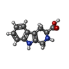


| #7: Chemical |  Type: L-peptide linking / Mass: 216.236 Da / Num. of mol.: 2 / Source method: obtained synthetically / Formula: C12H12N2O2 Type: L-peptide linking / Mass: 216.236 Da / Num. of mol.: 2 / Source method: obtained synthetically / Formula: C12H12N2O2#8: Chemical | Phosphate Mass: 94.971 Da / Num. of mol.: 2 / Source method: obtained synthetically / Formula: PO4 Mass: 94.971 Da / Num. of mol.: 2 / Source method: obtained synthetically / Formula: PO4#9: Chemical | ChemComp-CA /  Mass: 40.078 Da / Num. of mol.: 6 / Source method: obtained synthetically / Formula: Ca Mass: 40.078 Da / Num. of mol.: 6 / Source method: obtained synthetically / Formula: Ca#10: Chemical |  Mass: 303.826 Da / Num. of mol.: 2 / Source method: obtained synthetically / Formula: C18H22ClNO Mass: 303.826 Da / Num. of mol.: 2 / Source method: obtained synthetically / Formula: C18H22ClNO#11: Chemical | ChemComp-Y01 / |  Mass: 486.726 Da / Num. of mol.: 1 / Source method: obtained synthetically / Formula: C31H50O4 Mass: 486.726 Da / Num. of mol.: 1 / Source method: obtained synthetically / Formula: C31H50O4#12: Chemical | ChemComp-A1AF7 / ( | Mass: 749.007 Da / Num. of mol.: 1 / Source method: obtained synthetically / Formula: C40H77O10P |
|---|
-Details
| Has ligand of interest | N |
|---|
-Experimental details
-Experiment
| Experiment | Method: ELECTRON MICROSCOPY |
|---|---|
| EM experiment | Aggregation state: PARTICLE / 3D reconstruction method: single particle reconstruction |
- Sample preparation
| Component | Name: Human CaSR in complex with Gi3 protein / Type: COMPLEX / Entity ID: #1-#4 / Source: RECOMBINANT | |||||||||||||||||||||||||||||||||||
|---|---|---|---|---|---|---|---|---|---|---|---|---|---|---|---|---|---|---|---|---|---|---|---|---|---|---|---|---|---|---|---|---|---|---|---|---|
| Molecular weight | Value: 0.288 MDa / Experimental value: NO | |||||||||||||||||||||||||||||||||||
| Source (natural) | Organism: Homo sapiens (human) | |||||||||||||||||||||||||||||||||||
| Source (recombinant) | Organism: Homo sapiens (human) / Cell: HEK293 GnTI- | |||||||||||||||||||||||||||||||||||
| Buffer solution | pH: 7.5 | |||||||||||||||||||||||||||||||||||
| Buffer component |
| |||||||||||||||||||||||||||||||||||
| Specimen | Conc.: 3.5 mg/ml / Embedding applied: NO / Shadowing applied: NO / Staining applied: NO / Vitrification applied: YES | |||||||||||||||||||||||||||||||||||
| Specimen support | Grid material: GOLD / Grid mesh size: 300 divisions/in. / Grid type: Quantifoil R0.6/1 | |||||||||||||||||||||||||||||||||||
| Vitrification | Instrument: FEI VITROBOT MARK IV / Cryogen name: ETHANE / Humidity: 100 % / Chamber temperature: 277 K Details: The sample was blotted for 6s before plunge-frozen. |
- Electron microscopy imaging
| Experimental equipment |  Model: Titan Krios / Image courtesy: FEI Company |
|---|---|
| Microscopy | Model: FEI TITAN KRIOS |
| Electron gun | Electron source: FIELD EMISSION GUN / Accelerating voltage: 300 kV / Illumination mode: FLOOD BEAM |
| Electron lens | Mode: BRIGHT FIELDBright-field microscopy / Nominal magnification: 105000 X / Nominal defocus max: 1800 nm / Nominal defocus min: 1000 nm / Cs: 2.7 mm / C2 aperture diameter: 100 µm / Alignment procedure: COMA FREE |
| Specimen holder | Cryogen: NITROGEN / Specimen holder model: FEI TITAN KRIOS AUTOGRID HOLDER / Temperature (max): 100 K |
| Image recording | Average exposure time: 2.5 sec. / Electron dose: 70.14 e/Å2 / Film or detector model: GATAN K3 BIOQUANTUM (6k x 4k) / Num. of grids imaged: 2 / Num. of real images: 16504 |
| EM imaging optics | Energyfilter name: GIF Bioquantum / Energyfilter slit width: 20 eV |
| Image scans | Width: 5760 / Height: 4092 |
- Processing
| EM software |
| ||||||||||||||||||||||||||||||||||||
|---|---|---|---|---|---|---|---|---|---|---|---|---|---|---|---|---|---|---|---|---|---|---|---|---|---|---|---|---|---|---|---|---|---|---|---|---|---|
| CTF correction | Type: PHASE FLIPPING AND AMPLITUDE CORRECTION | ||||||||||||||||||||||||||||||||||||
| Particle selection | Num. of particles selected: 2792317 | ||||||||||||||||||||||||||||||||||||
| Symmetry | Point symmetry: C1 (asymmetric) | ||||||||||||||||||||||||||||||||||||
| 3D reconstruction | Resolution: 3.8 Å / Resolution method: FSC 0.143 CUT-OFF / Num. of particles: 55985 / Symmetry type: POINT | ||||||||||||||||||||||||||||||||||||
| Atomic model building | Protocol: FLEXIBLE FIT / Space: REAL | ||||||||||||||||||||||||||||||||||||
| Atomic model building | 3D fitting-ID: 1 / Source name: PDB / Type: experimental model
| ||||||||||||||||||||||||||||||||||||
| Refine LS restraints |
|