Movie
Movie Controller

[English] 日本語
 Yorodumi
- PDB-7sil: Structure of positive allosteric modulator-bound active human cal... -
+ Open data

- Basic information
| Entry | Database: PDB / ID: 7sil | |||||||||||||||||||||
|---|---|---|---|---|---|---|---|---|---|---|---|---|---|---|---|---|---|---|---|---|---|---|
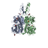
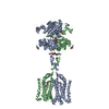
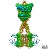
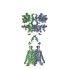
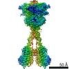
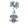
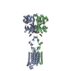
| Title | Structure of positive allosteric modulator-bound active human calcium-sensing receptor | |||||||||||||||||||||
 Components Components | Isoform 1 of Extracellular calcium-sensing receptor | |||||||||||||||||||||
 Keywords Keywords |  MEMBRANE PROTEIN / calcium-sensing receptor / cryo-EM structure / allosteric modulation / activation mechanism / symmetry MEMBRANE PROTEIN / calcium-sensing receptor / cryo-EM structure / allosteric modulation / activation mechanism / symmetry | |||||||||||||||||||||
| Function / homology | Chem-9IG / CHOLESTEROL / PHOSPHATE ION / CYCLOMETHYLTRYPTOPHAN / Isoform 1 of Extracellular calcium-sensing receptor Function and homology information Function and homology information | |||||||||||||||||||||
| Biological species |  Homo sapiens (human) Homo sapiens (human) | |||||||||||||||||||||
| Method | ELECTRON MICROSCOPY / single particle reconstruction / cryo EM / Resolution: 2.7 Å | |||||||||||||||||||||
 Authors Authors | Park, J. / Zuo, H. / Frangaj, A. / Fu, Z. / Yen, L.Y. / Zhang, Z. / Mosyak, L. / Slavkovich, V.N. / Liu, J. / Ray, K.M. ...Park, J. / Zuo, H. / Frangaj, A. / Fu, Z. / Yen, L.Y. / Zhang, Z. / Mosyak, L. / Slavkovich, V.N. / Liu, J. / Ray, K.M. / Cao, B. / Vallese, F. / Geng, Y. / Chen, S. / Grassucci, R. / Dandey, V.P. / Tan, Y.Z. / Eng, E. / Lee, Y. / Kloss, B. / Liu, Z. / Hendrickson, W.A. / Potter, C.S. / Carragher, B. / Graziano, J. / Conigrave, A.D. / Frank, J. / Clarke, O.B. / Fan, Q.R. | |||||||||||||||||||||
| Funding support |  United States, 6items United States, 6items
| |||||||||||||||||||||
 Citation Citation | Journal: Proc Natl Acad Sci U S A / Year: 2021 Title: Symmetric activation and modulation of the human calcium-sensing receptor. Authors: Jinseo Park / Hao Zuo / Aurel Frangaj / Ziao Fu / Laura Y Yen / Zhening Zhang / Lidia Mosyak / Vesna N Slavkovich / Jonathan Liu / Kimberly M Ray / Baohua Cao / Francesca Vallese / Yong Geng ...Authors: Jinseo Park / Hao Zuo / Aurel Frangaj / Ziao Fu / Laura Y Yen / Zhening Zhang / Lidia Mosyak / Vesna N Slavkovich / Jonathan Liu / Kimberly M Ray / Baohua Cao / Francesca Vallese / Yong Geng / Shaoxia Chen / Robert Grassucci / Venkata P Dandey / Yong Zi Tan / Edward Eng / Yeji Lee / Brian Kloss / Zheng Liu / Wayne A Hendrickson / Clinton S Potter / Bridget Carragher / Joseph Graziano / Arthur D Conigrave / Joachim Frank / Oliver B Clarke / Qing R Fan /    Abstract: The human extracellular calcium-sensing (CaS) receptor controls plasma Ca levels and contributes to nutrient-dependent maintenance and metabolism of diverse organs. Allosteric modulation of the CaS ...The human extracellular calcium-sensing (CaS) receptor controls plasma Ca levels and contributes to nutrient-dependent maintenance and metabolism of diverse organs. Allosteric modulation of the CaS receptor corrects disorders of calcium homeostasis. Here, we report the cryogenic-electron microscopy reconstructions of a near-full-length CaS receptor in the absence and presence of allosteric modulators. Activation of the homodimeric CaS receptor requires a break in the transmembrane 6 (TM6) helix of each subunit, which facilitates the formation of a TM6-mediated homodimer interface and expansion of homodimer interactions. This transformation in TM6 occurs without a positive allosteric modulator. Two modulators with opposite functional roles bind to overlapping sites within the transmembrane domain through common interactions, acting to stabilize distinct rotamer conformations of key residues on the TM6 helix. The positive modulator reinforces TM6 distortion and maximizes subunit contact to enhance receptor activity, while the negative modulator strengthens an intact TM6 to dampen receptor function. In both active and inactive states, the receptor displays symmetrical transmembrane conformations that are consistent with its homodimeric assembly. | |||||||||||||||||||||
| History |
|
- Structure visualization
| Movie |
Movie viewer |
|---|---|
| Structure viewer | Molecule: MolmilJmol/JSmol |
- Downloads & links
-Download
| PDBx/mmCIF format | 7sil.cif.gz | 288.8 KB | Display | PDBx/mmCIF format |
|---|---|---|---|---|
| PDB format | pdb7sil.ent.gz | 242.5 KB | Display | PDB format |
| PDBx/mmJSON format | 7sil.json.gz | Tree view | PDBx/mmJSON format | |
| Others |  Other downloads Other downloads |
-Validation report
| Arichive directory | https://data.pdbj.org/pub/pdb/validation_reports/si/7silftp://data.pdbj.org/pub/pdb/validation_reports/si/7sil | HTTPS FTP |
|---|
-Related structure data
| Related structure data |  25143MC 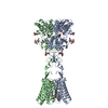 7simC 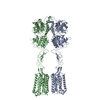 7sinC M: map data used to model this data C: citing same article ( |
|---|---|
| Similar structure data | |
| EM raw data | EMPIAR-10834 (Title: Structures of positive allosteric modulator-bound and unbound active human calcium-sensing receptor Data size: 3.8 TB Data #1: Unaligned multi-frame micrographs of calcium-sensing receptor in both PAM-bound and unbound active states [micrographs - multiframe]) |


-Links
 PDBj
PDBj


- Assembly
| Deposited unit | 
|
|---|---|
| 1 |
|
-Components
-Protein , 1 types, 2 molecules AB
| #1: Protein | Mass: 99299.461 Da / Num. of mol.: 2 Source method: isolated from a genetically manipulated source Source: (gene. exp.) Homo sapiens (human) / Gene: CASR, GPRC2A, PCAR1 / Cell line (production host): HEK293 GnTl- / Production host: Homo sapiens (human) / References: UniProt: P41180-1 |
|---|
-Sugars , 2 types, 10 molecules 
| #2: Polysaccharide | 2-acetamido-2-deoxy-beta-D-glucopyranose-(1-4)-2-acetamido-2-deoxy-beta-D-glucopyranose / Mass: 424.401 Da / Num. of mol.: 4Source method: isolated from a genetically manipulated source #3: Sugar | ChemComp-NAG / N-Acetylglucosamine Type: D-saccharide, beta linking / Mass: 221.208 Da / Num. of mol.: 6 / Source method: obtained synthetically / Formula: C8H15NO6 Type: D-saccharide, beta linking / Mass: 221.208 Da / Num. of mol.: 6 / Source method: obtained synthetically / Formula: C8H15NO6 |
|---|
-Non-polymers , 5 types, 22 molecules 


| #4: Chemical |  Type: L-peptide linking / Mass: 216.236 Da / Num. of mol.: 2 / Source method: obtained synthetically / Formula: C12H12N2O2 / Feature type: SUBJECT OF INVESTIGATION Type: L-peptide linking / Mass: 216.236 Da / Num. of mol.: 2 / Source method: obtained synthetically / Formula: C12H12N2O2 / Feature type: SUBJECT OF INVESTIGATION#5: Chemical | Phosphate Mass: 94.971 Da / Num. of mol.: 2 / Source method: obtained synthetically / Formula: PO4 / Feature type: SUBJECT OF INVESTIGATION Mass: 94.971 Da / Num. of mol.: 2 / Source method: obtained synthetically / Formula: PO4 / Feature type: SUBJECT OF INVESTIGATION#6: Chemical | ChemComp-CA /  Mass: 40.078 Da / Num. of mol.: 8 / Source method: obtained synthetically / Formula: Ca / Feature type: SUBJECT OF INVESTIGATION Mass: 40.078 Da / Num. of mol.: 8 / Source method: obtained synthetically / Formula: Ca / Feature type: SUBJECT OF INVESTIGATION#7: Chemical |  Mass: 303.826 Da / Num. of mol.: 2 / Source method: obtained synthetically / Formula: C18H22ClNO / Feature type: SUBJECT OF INVESTIGATION Mass: 303.826 Da / Num. of mol.: 2 / Source method: obtained synthetically / Formula: C18H22ClNO / Feature type: SUBJECT OF INVESTIGATION#8: Chemical | ChemComp-CLR / Cholesterol Mass: 386.654 Da / Num. of mol.: 8 / Source method: obtained synthetically / Formula: C27H46O Mass: 386.654 Da / Num. of mol.: 8 / Source method: obtained synthetically / Formula: C27H46O |
|---|
-Details
| Has ligand of interest | Y |
|---|
-Experimental details
-Experiment
| Experiment | Method: ELECTRON MICROSCOPY |
|---|---|
| EM experiment | Aggregation state: PARTICLE / 3D reconstruction method: single particle reconstruction |
- Sample preparation
| Component | Name: Homodimer human calcium sensing receptor / Type: COMPLEX / Entity ID: #1 / Source: RECOMBINANT | ||||||||||||||||||||||||||||||||||||||||
|---|---|---|---|---|---|---|---|---|---|---|---|---|---|---|---|---|---|---|---|---|---|---|---|---|---|---|---|---|---|---|---|---|---|---|---|---|---|---|---|---|---|
| Molecular weight | Value: 0.192 MDa / Experimental value: NO | ||||||||||||||||||||||||||||||||||||||||
| Source (natural) | Organism: Homo sapiens (human) | ||||||||||||||||||||||||||||||||||||||||
| Source (recombinant) | Organism: Homo sapiens (human) / Cell: HEK 293 GnTl- / Plasmid: modified bacMam | ||||||||||||||||||||||||||||||||||||||||
| Buffer solution | pH: 7.5 Details: Solution were made fresh from concentrated, and filtered to avoid microbial contamination. | ||||||||||||||||||||||||||||||||||||||||
| Buffer component |
| ||||||||||||||||||||||||||||||||||||||||
| Specimen | Conc.: 2.6 mg/ml / Embedding applied: NO / Shadowing applied: NO / Staining applied: NO / Vitrification applied: YES / Details: This sample was monodisperse | ||||||||||||||||||||||||||||||||||||||||
| Specimen support | Grid material: GOLD / Grid mesh size: 300 divisions/in. / Grid type: Quantifoil R0.6/1 | ||||||||||||||||||||||||||||||||||||||||
| Vitrification | Instrument: FEI VITROBOT MARK IV / Cryogen name: ETHANE / Humidity: 100 % / Chamber temperature: 277 K / Details: blot for 4 seconds before plunging |
- Electron microscopy imaging
| Experimental equipment |  Model: Titan Krios / Image courtesy: FEI Company |
|---|---|
| Microscopy | Model: FEI TITAN KRIOS / Details: Preliminary grid screening was performed manually. |
| Electron gun | Electron source: FIELD EMISSION GUN / Accelerating voltage: 300 kV / Illumination mode: FLOOD BEAM |
| Electron lens | Mode: BRIGHT FIELDBright-field microscopy / Nominal magnification: 105000 X / Nominal defocus max: 2500 nm / Nominal defocus min: 1000 nm / Cs: 2.7 mm / C2 aperture diameter: 70 µm / Alignment procedure: COMA FREE |
| Specimen holder | Cryogen: NITROGEN / Specimen holder model: FEI TITAN KRIOS AUTOGRID HOLDER / Temperature (max): 100 K |
| Image recording | Electron dose: 70.35 e/Å2 / Film or detector model: GATAN K3 (6k x 4k) / Num. of grids imaged: 1 / Num. of real images: 13246 |
| EM imaging optics | Energyfilter name: GIF Quantum ER / Energyfilter slit width: 20 eV |
- Processing
| Software | Name: PHENIX / Version: 1.19.1_4122: / Classification: refinement | ||||||||||||||||||||||||||||||||||||||||||||||||||
|---|---|---|---|---|---|---|---|---|---|---|---|---|---|---|---|---|---|---|---|---|---|---|---|---|---|---|---|---|---|---|---|---|---|---|---|---|---|---|---|---|---|---|---|---|---|---|---|---|---|---|---|
| EM software |
| ||||||||||||||||||||||||||||||||||||||||||||||||||
| CTF correction | Type: PHASE FLIPPING AND AMPLITUDE CORRECTION | ||||||||||||||||||||||||||||||||||||||||||||||||||
| Particle selection | Num. of particles selected: 892000 | ||||||||||||||||||||||||||||||||||||||||||||||||||
| Symmetry | Point symmetry: C2 (2 fold cyclic) | ||||||||||||||||||||||||||||||||||||||||||||||||||
| 3D reconstruction | Resolution: 2.7 Å / Resolution method: FSC 0.143 CUT-OFF / Num. of particles: 130000 / Algorithm: FOURIER SPACE / Details: Global(ECD) 2.7A TM 3.4A / Num. of class averages: 1 / Symmetry type: POINT | ||||||||||||||||||||||||||||||||||||||||||||||||||
| Atomic model building | Protocol: FLEXIBLE FIT / Space: REAL | ||||||||||||||||||||||||||||||||||||||||||||||||||
| Atomic model building |
| ||||||||||||||||||||||||||||||||||||||||||||||||||
| Refine LS restraints |
|