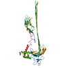
 ムービー
ムービー コントローラー
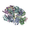
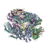
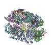
 構造ビューア
 EMN検索について


-検索条件
-検索結果
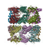
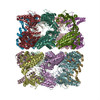
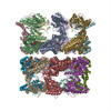
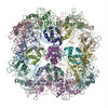
検索 (著者・登録者: h. & r. & saibil)の結果53件中、1から50件目までを表示しています
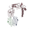
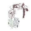
PDB-8rqm: 
Alpha-synuclein amyloid fibrils
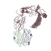
PDB-8rrr:
Alpha-synuclein amyloid fibril

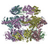
PDB-8ba8:
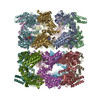
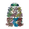
CryoEM structure of GroEL-ADP.BeF3-Rubisco.

PDB-8ba9:
CryoEM structure of GroEL-GroES-ADP.AlF3-Rubisco.

PDB-8ba7:
CryoEM structure of nucleotide-free GroEL-Rubisco.
PDB-8b6v:
Mp2Ba1 pre-pore

PDB-8b6w:
Mpf2Ba1 pore

PDB-8a00:
Infectious mouse-adapted ME7 scrapie prion fibril purified from terminally-infected mouse brains
PDB-7qig:
Infectious mouse-adapted RML scrapie prion fibril purified from terminally-infected mouse brains
PDB-7pag:
The pore conformation of lymphocyte perforin
PDB-6qs4:
Two-Step Activation Mechanism of the ClpB Disaggregase for Sequential Substrate Threading by the Main ATPase Motor.
PDB-6qs6:
ClpB (DWB and K476C mutant) bound to casein in presence of ATPgammaS - state KC-1
PDB-6qs7:
ClpB (DWB and K476C mutant) bound to casein in presence of ATPgammaS - state KC-2A
PDB-6qs8:
ClpB (DWB and K476C mutant) bound to casein in presence of ATPgammaS - state KC-2B
PDB-6rn2:
ClpB (DWB mutant) bound to casein in presence of ATPgammaS - state WT-1
PDB-6rn3:
ClpB (DWB mutant) bound to casein in presence of ATPgammaS - state WT-2A
PDB-6rn4:
ClpB (DWB mutant) bound to casein in presence of ATPgammaS - state WT-2B
PDB-5ofo:
Cryo EM structure of the E. coli disaggregase ClpB (BAP form, DWB mutant), in the ATPgammaS state, bound to the model substrate casein
PDB-5og1:
Cryo EM structure of the E. coli disaggregase ClpB (BAP form, DWB mutant), in the ATPgammaS state
PDB-5fmw:
The poly-C9 component of the Complement Membrane Attack Complex
PDB-4v2t:
Membrane embedded pleurotolysin pore with 13 fold symmetry
PDB-4v3a:
Membrane bound pleurotolysin prepore (TMH1 lock) trapped with engineered disulphide cross-link
PDB-4v3m:
Membrane bound pleurotolysin prepore (TMH2 helix lock) trapped with engineered disulphide cross-link
PDB-4v3n:
Membrane bound pleurotolysin prepore (TMH2 strand lock) trapped with engineered disulphide cross-link
PDB-4d2q:
Negative-stain electron microscopy of E. coli ClpB mutant E432A (BAP form bound to ClpP)
PDB-4d2u:
Negative-stain electron microscopy of E. coli ClpB (BAP form bound to ClpP)
PDB-4d2x:
Negative-stain electron microscopy of E. coli ClpB of Y503D hyperactive mutant (BAP form bound to ClpP)
PDB-2m5k:
Atomic-resolution structure of a doublet cross-beta amyloid fibril
PDB-2m5m:
Atomic-resolution structure of a triplet cross-beta amyloid fibril
PDB-3zpk:
Atomic-resolution structure of a quadruplet cross-beta amyloid fibril.
PDB-4aaq:
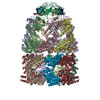
ATP-triggered molecular mechanics of the chaperonin GroEL
PDB-4aar:
ATP-triggered molecular mechanics of the chaperonin GroEL
PDB-4aas:
ATP-triggered molecular mechanics of the chaperonin GroEL
PDB-4aau:
ATP-triggered molecular mechanics of the chaperonin GroEL
PDB-4ab2:
ATP-triggered molecular mechanics of the chaperonin GroEL
PDB-4ab3:
ATP-triggered molecular mechanics of the chaperonin GroEL
PDB-4a8b:
Symmetrized cryo-EM reconstruction of E. coli DegQ 12-mer in complex with lysozymes
PDB-4a8c:
Symmetrized cryo-EM reconstruction of E. coli DegQ 12-mer in complex with a binding peptide
PDB-4a8d:
DegP dodecamer with bound OMP
PDB-4a9g:
Symmetrized cryo-EM reconstruction of E. coli DegQ 24-mer in complex with beta-casein
PDB-4a8a:
Asymmetric cryo-EM reconstruction of E. coli DegQ 12-mer in complex with lysozyme
PDB-2zle:
Cryo-EM structure of DegP12/OMP
PDB-2h50:
Multiple distinct assemblies reveal conformational flexibility in the small heat shock protein Hsp26
PDB-2h53:
Multiple distinct assemblies reveal conformational flexibility in the small heat shock protein Hsp26
PDB-2cgt:
GROEL-ADP-gp31 COMPLEX
PDB-2c7e:
REVISED ATOMIC STRUCTURE FITTING INTO A GROEL(D398A)-ATP7 CRYO-EM MAP (EMD 1047)
PDB-2c7c:
FITTED COORDINATES FOR GROEL-ATP7-GROES CRYO-EM COMPLEX (EMD-1180)
PDB-2c7d:
Fitted coordinates for GroEL-ADP7-GroES Cryo-EM complex (EMD-1181)

PDB-2byu:
Negative stain EM reconstruction of M.tuberculosis Acr1(Hsp 16.3) fitted with wheat sHSP dimer
PDB-2bk1:
The pore structure of pneumolysin, obtained by fitting the alpha carbon trace of perfringolysin O into a cryo-EM map
ページ:
 wwPDBはEMDBデータモデルのバージョン3へ移行します
