 Movie
Movie Controller


+ Open data

- Basic information
| Entry | Database: PDB / ID: 8ba9 | ||||||
|---|---|---|---|---|---|---|---|
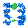
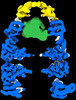
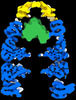
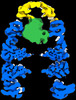
| Title | CryoEM structure of GroEL-GroES-ADP.AlF3-Rubisco. | ||||||
 Components Components |
| ||||||
 Keywords Keywords | CHAPERONE / GroEL / GroES | ||||||
| Function / homology |  Function and homology information Function and homology informationGroEL-GroES complex / chaperonin ATPase / virion assembly / chaperone cofactor-dependent protein refolding / protein folding chaperone / isomerase activity / ATP-dependent protein folding chaperone / response to radiation / unfolded protein binding / protein folding ...GroEL-GroES complex / chaperonin ATPase / virion assembly / chaperone cofactor-dependent protein refolding / protein folding chaperone / isomerase activity / ATP-dependent protein folding chaperone / response to radiation / unfolded protein binding / protein folding / protein-folding chaperone binding / protein refolding / response to heat / magnesium ion binding / ATP hydrolysis activity / ATP binding / identical protein binding / membrane / metal ion binding / cytosol Similarity search - Function | ||||||
| Biological species |  | ||||||
| Method | ELECTRON MICROSCOPY / single particle reconstruction / cryo EM / Resolution: 3.7 Å | ||||||
 Authors Authors | Gardner, S. / Saibil, H.R. | ||||||
| Funding support |  United Kingdom, 1items United Kingdom, 1items
| ||||||
 Citation Citation | Journal: Proc Natl Acad Sci U S A / Year: 2023 Title: Structural basis of substrate progression through the bacterial chaperonin cycle. Authors: Scott Gardner / Michele C Darrow / Natalya Lukoyanova / Konstantinos Thalassinos / Helen R Saibil / Abstract: The bacterial chaperonin GroEL-GroES promotes protein folding through ATP-regulated cycles of substrate protein binding, encapsulation, and release. Here, we have used cryoEM to determine structures ...The bacterial chaperonin GroEL-GroES promotes protein folding through ATP-regulated cycles of substrate protein binding, encapsulation, and release. Here, we have used cryoEM to determine structures of GroEL, GroEL-ADP·BeF, and GroEL-ADP·AlF-GroES all complexed with the model substrate Rubisco. Our structures provide a series of snapshots that show how the conformation and interactions of non-native Rubisco change as it proceeds through the GroEL-GroES reaction cycle. We observe specific charged and hydrophobic GroEL residues forming strong initial contacts with non-native Rubisco. Binding of ATP or ADP·BeF to GroEL-Rubisco results in the formation of an intermediate GroEL complex displaying striking asymmetry in the ATP/ADP·BeF-bound ring. In this ring, four GroEL subunits bind Rubisco and the other three are in the GroES-accepting conformation, suggesting how GroEL can recruit GroES without releasing bound substrate. Our cryoEM structures of stalled GroEL-ADP·AlF-Rubisco-GroES complexes show Rubisco folding intermediates interacting with GroEL-GroES via different sets of residues. | ||||||
| History |
|
- Structure visualization
| Structure viewer | Molecule: MolmilJmol/JSmol |
|---|
- Downloads & links
-Download
| PDBx/mmCIF format | 8ba9.cif.gz | 2.4 MB | Display | PDBx/mmCIF format |
|---|---|---|---|---|
| PDB format | pdb8ba9.ent.gz | Display | PDB format | |
| PDBx/mmJSON format | 8ba9.json.gz | Tree view | PDBx/mmJSON format | |
| Others |  Other downloads Other downloads |
-Validation report
| Summary document | 8ba9_validation.pdf.gz | 2.3 MB | Display | wwPDB validaton report |
|---|---|---|---|---|
| Full document | 8ba9_full_validation.pdf.gz | 2.4 MB | Display | |
| Data in XML | 8ba9_validation.xml.gz | 207.1 KB | Display | |
| Data in CIF | 8ba9_validation.cif.gz | 314.7 KB | Display | |
| Arichive directory | https://data.pdbj.org/pub/pdb/validation_reports/ba/8ba9ftp://data.pdbj.org/pub/pdb/validation_reports/ba/8ba9 | HTTPS FTP |
-Related structure data
| Related structure data |  15942MC  8ba7C  8ba8C M: map data used to model this data C: citing same article ( |
|---|---|
| Similar structure data |
-Links
 PDBj
PDBj


- Assembly
| Deposited unit | 
|
|---|---|
| 1 |
|
-Components
-Protein , 2 types, 21 molecules ABCDEFGHIJKLMNOPQRSTU
| #1: Protein | Mass: 55220.105 Da / Num. of mol.: 14 Source method: isolated from a genetically manipulated source Source: (gene. exp.) #2: Protein | Mass: 10400.938 Da / Num. of mol.: 7 Source method: isolated from a genetically manipulated source Source: (gene. exp.) |
|---|
-Non-polymers , 4 types, 42 molecules 


| #3: Chemical | ChemComp-AF3 /  Mass: 83.977 Da / Num. of mol.: 7 / Source method: obtained synthetically / Formula: AlF3 Mass: 83.977 Da / Num. of mol.: 7 / Source method: obtained synthetically / Formula: AlF3#4: Chemical | ChemComp-ADP /  Mass: 427.201 Da / Num. of mol.: 14 / Source method: obtained synthetically / Formula: C10H15N5O10P2 / Comment: ADP, energy-carrying molecule*YM Mass: 427.201 Da / Num. of mol.: 14 / Source method: obtained synthetically / Formula: C10H15N5O10P2 / Comment: ADP, energy-carrying molecule*YM#5: Chemical | ChemComp-MG /  Mass: 24.305 Da / Num. of mol.: 14 / Source method: obtained synthetically / Formula: Mg Mass: 24.305 Da / Num. of mol.: 14 / Source method: obtained synthetically / Formula: Mg#6: Chemical | ChemComp-K /  Mass: 39.098 Da / Num. of mol.: 7 / Source method: obtained synthetically / Formula: K Mass: 39.098 Da / Num. of mol.: 7 / Source method: obtained synthetically / Formula: K |
|---|
-Details
| Has ligand of interest | N |
|---|
-Experimental details
-Experiment
| Experiment | Method: ELECTRON MICROSCOPY |
|---|---|
| EM experiment | Aggregation state: PARTICLE / 3D reconstruction method: single particle reconstruction |
- Sample preparation
| Component | Name: GroEL-ADP.BeFx-Rubisco / Type: COMPLEX / Entity ID: #1 / Source: RECOMBINANT |
|---|---|
| Molecular weight | Value: 0.802 MDa / Experimental value: NO |
| Source (natural) | Organism: |
| Source (recombinant) | Organism: |
| Buffer solution | pH: 7.5 |
| Specimen | Conc.: 2.7 mg/ml / Embedding applied: NO / Shadowing applied: NO / Staining applied: NO / Vitrification applied: YES |
| Vitrification | Instrument: SPOTITON / Cryogen name: ETHANE / Humidity: 80 % / Chamber temperature: 293 K Details: The grid was prepared using a chameleon (SPT Labtech). |
- Electron microscopy imaging
| Experimental equipment |  Model: Titan Krios / Image courtesy: FEI Company |
|---|---|
| Microscopy | Model: FEI TITAN KRIOS |
| Electron gun | Electron source:  FIELD EMISSION GUN / Accelerating voltage: 300 kV / Illumination mode: SPOT SCAN FIELD EMISSION GUN / Accelerating voltage: 300 kV / Illumination mode: SPOT SCAN |
| Electron lens | Mode: BRIGHT FIELD / Nominal defocus max: 3000 nm / Nominal defocus min: 1400 nm / Cs: 2.7 mm / C2 aperture diameter: 70 µm |
| Image recording | Electron dose: 40.2 e/Å2 / Film or detector model: GATAN K3 BIOQUANTUM (6k x 4k) / Num. of grids imaged: 2 |
- Processing
| EM software |
| ||||||||||||||||||||
|---|---|---|---|---|---|---|---|---|---|---|---|---|---|---|---|---|---|---|---|---|---|
| CTF correction | Type: PHASE FLIPPING AND AMPLITUDE CORRECTION | ||||||||||||||||||||
| Particle selection | Num. of particles selected: 7712622 | ||||||||||||||||||||
| 3D reconstruction | Resolution: 3.7 Å / Resolution method: FSC 0.143 CUT-OFF / Num. of particles: 30965 / Num. of class averages: 1 / Symmetry type: POINT |
