 Movie
Movie Controller


+ Open data

- Basic information
| Entry | Database: PDB / ID: 7pag | |||||||||||||||||||||
|---|---|---|---|---|---|---|---|---|---|---|---|---|---|---|---|---|---|---|---|---|---|---|
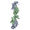
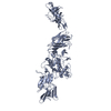
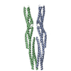
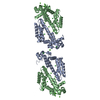
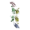
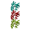
| Title | The pore conformation of lymphocyte perforin | |||||||||||||||||||||
 Components Components | Perforin-1 | |||||||||||||||||||||
 Keywords Keywords | IMMUNE SYSTEM / Pore forming protein perforin | |||||||||||||||||||||
| Function / homology |  Function and homology information Function and homology informationimmune response to tumor cell / granzyme-mediated programmed cell death signaling pathway / cytolytic granule / pore-forming activity / protein transmembrane transport / immunological synapse formation / wide pore channel activity / protein import / positive regulation of killing of cells of another organism / defense response to tumor cell ...immune response to tumor cell / granzyme-mediated programmed cell death signaling pathway / cytolytic granule / pore-forming activity / protein transmembrane transport / immunological synapse formation / wide pore channel activity / protein import / positive regulation of killing of cells of another organism / defense response to tumor cell / immunological synapse / protein secretion / endosome lumen / protein homooligomerization / T cell mediated cytotoxicity / circadian rhythm / cytoplasmic vesicle / defense response to virus / killing of cells of another organism / calcium ion binding / extracellular space / membrane / identical protein binding / plasma membrane / cytosolSimilarity search - Function | |||||||||||||||||||||
| Biological species |  Mus musculus (house mouse) Mus musculus (house mouse) | |||||||||||||||||||||
| Method | ELECTRON MICROSCOPY / single particle reconstruction / cryo EM / Resolution: 4 Å | |||||||||||||||||||||
 Authors Authors | Ivanova, M.E. / Lukoyanova, N. / Malhotra, S. / Topf, M. / Trapani, J.A. / Voskoboinik, I. / Saibil, H.R. | |||||||||||||||||||||
| Funding support |  United Kingdom, 6items United Kingdom, 6items
| |||||||||||||||||||||
 Citation Citation | Journal: Sci Adv / Year: 2022 Title: The pore conformation of lymphocyte perforin. Authors: Marina E Ivanova / Natalya Lukoyanova / Sony Malhotra / Maya Topf / Joseph A Trapani / Ilia Voskoboinik / Helen R Saibil /   Abstract: Perforin is a pore-forming protein that facilitates rapid killing of pathogen-infected or cancerous cells by the immune system. Perforin is released from cytotoxic lymphocytes, together with ...Perforin is a pore-forming protein that facilitates rapid killing of pathogen-infected or cancerous cells by the immune system. Perforin is released from cytotoxic lymphocytes, together with proapoptotic granzymes, to bind to a target cell membrane where it oligomerizes and forms pores. The pores allow granzyme entry, which rapidly triggers the apoptotic death of the target cell. Here, we present a 4-Å resolution cryo-electron microscopy structure of the perforin pore, revealing previously unidentified inter- and intramolecular interactions stabilizing the assembly. During pore formation, the helix-turn-helix motif moves away from the bend in the central β sheet to form an intermolecular contact. Cryo-electron tomography shows that prepores form on the membrane surface with minimal conformational changes. Our findings suggest the sequence of conformational changes underlying oligomerization and membrane insertion, and explain how several pathogenic mutations affect function. | |||||||||||||||||||||
| History |
|
- Structure visualization
| Movie |
Movie viewer |
|---|---|
| Structure viewer | Molecule: MolmilJmol/JSmol |
- Downloads & links
-Download
| PDBx/mmCIF format | 7pag.cif.gz | 108.4 KB | Display | PDBx/mmCIF format |
|---|---|---|---|---|
| PDB format | pdb7pag.ent.gz | 85 KB | Display | PDB format |
| PDBx/mmJSON format | 7pag.json.gz | Tree view | PDBx/mmJSON format | |
| Others |  Other downloads Other downloads |
-Validation report
| Arichive directory | https://data.pdbj.org/pub/pdb/validation_reports/pa/7pagftp://data.pdbj.org/pub/pdb/validation_reports/pa/7pag | HTTPS FTP |
|---|
-Related structure data
| Related structure data | 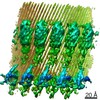 13269MC M: map data used to model this data C: citing same article ( |
|---|---|
| Similar structure data |

-Links
 PDBj
PDBj


- Assembly
| Deposited unit | 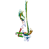
|
|---|---|
| 1 |
|
-Components
| #1: Protein | / P1 / Cytolysin / Lymphocyte pore-forming protein Mass: 60827.051 Da / Num. of mol.: 1 Source method: isolated from a genetically manipulated source Source: (gene. exp.) Mus musculus (house mouse) / Gene: Prf1, Pfp / Production host:   Spodoptera frugiperda (fall armyworm) / References: UniProt: P10820 Spodoptera frugiperda (fall armyworm) / References: UniProt: P10820 | ||||
|---|---|---|---|---|---|
| #2: Chemical |   Mass: 40.078 Da / Num. of mol.: 3 / Source method: obtained synthetically / Formula: Ca Mass: 40.078 Da / Num. of mol.: 3 / Source method: obtained synthetically / Formula: Ca#3: Sugar | ChemComp-NAG / | N-Acetylglucosamine  Type: D-saccharide, beta linking / Mass: 221.208 Da / Num. of mol.: 1 Type: D-saccharide, beta linking / Mass: 221.208 Da / Num. of mol.: 1Source method: isolated from a genetically manipulated source Formula: C8H15NO6 Has ligand of interest | N | |
-Experimental details
-Experiment
| Experiment | Method: ELECTRON MICROSCOPY |
|---|---|
| EM experiment | Aggregation state: PARTICLE / 3D reconstruction method: single particle reconstruction |
- Sample preparation
| Component | Name: Oligomerised mouse Perforin in its inserted state / Type: COMPLEX / Entity ID: #1 / Source: RECOMBINANT |
|---|---|
| Molecular weight | Experimental value: YES |
| Source (natural) | Organism: Mus musculus (house mouse) |
| Source (recombinant) | Organism: Spodoptera frugiperda (fall armyworm) |
| Buffer solution | pH: 7.5 |
| Specimen | Embedding applied: NO / Shadowing applied: NO / Staining applied: NO / Vitrification applied: YES |
| Specimen support | Grid material: GOLD / Grid type: UltrAuFoil R2/2 |
| Vitrification | Cryogen name: ETHANE |
- Electron microscopy imaging
| Experimental equipment |  Model: Titan Krios / Image courtesy: FEI Company |
|---|---|
| Microscopy | Model: FEI TITAN KRIOS |
| Electron gun | Electron source: FIELD EMISSION GUN / Accelerating voltage: 300 kV / Illumination mode: FLOOD BEAM |
| Electron lens | Mode: BRIGHT FIELDBright-field microscopy |
| Image recording | Electron dose: 49.6 e/Å2 / Film or detector model: GATAN K3 BIOQUANTUM (6k x 4k) |
- Processing
| CTF correction | Type: PHASE FLIPPING AND AMPLITUDE CORRECTION |
|---|---|
| 3D reconstruction | Resolution: 4 Å / Resolution method: FSC 0.143 CUT-OFF / Num. of particles: 229789 Details: 229,789 particles were symmetry expanded with C22 symmetry giving the final number of particles 5,055,358 Symmetry type: POINT |
