 Movie
Movie Controller


+ Open data

- Basic information
| Entry | Database: EMDB / ID: EMD-13269 | |||||||||||||||||||||
|---|---|---|---|---|---|---|---|---|---|---|---|---|---|---|---|---|---|---|---|---|---|---|
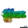
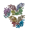
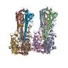
| Title | The pore conformation of lymphocyte perforin | |||||||||||||||||||||
 Map data Map data | ||||||||||||||||||||||
 Sample Sample |
| |||||||||||||||||||||
| Function / homology |  Function and homology information Function and homology informationimmune response to tumor cell / granzyme-mediated programmed cell death signaling pathway /  cytolytic granule / pore-forming activity / protein transmembrane transport / immunological synapse formation / wide pore channel activity / protein import / positive regulation of killing of cells of another organism / defense response to tumor cell ...immune response to tumor cell / granzyme-mediated programmed cell death signaling pathway / cytolytic granule / pore-forming activity / protein transmembrane transport / immunological synapse formation / wide pore channel activity / protein import / positive regulation of killing of cells of another organism / defense response to tumor cell / immunological synapse / protein secretion / endosome lumen / protein homooligomerization / T cell mediated cytotoxicity / circadian rhythm / cytoplasmic vesicle / defense response to virus / killing of cells of another organism / calcium ion binding / extracellular space / membrane / identical protein binding / plasma membrane / cytosol cytolytic granule / pore-forming activity / protein transmembrane transport / immunological synapse formation / wide pore channel activity / protein import / positive regulation of killing of cells of another organism / defense response to tumor cell ...immune response to tumor cell / granzyme-mediated programmed cell death signaling pathway / cytolytic granule / pore-forming activity / protein transmembrane transport / immunological synapse formation / wide pore channel activity / protein import / positive regulation of killing of cells of another organism / defense response to tumor cell / immunological synapse / protein secretion / endosome lumen / protein homooligomerization / T cell mediated cytotoxicity / circadian rhythm / cytoplasmic vesicle / defense response to virus / killing of cells of another organism / calcium ion binding / extracellular space / membrane / identical protein binding / plasma membrane / cytosolSimilarity search - Function | |||||||||||||||||||||
| Biological species |  Mus musculus (house mouse) Mus musculus (house mouse) | |||||||||||||||||||||
| Method | single particle reconstruction / cryo EM / Resolution: 4.0 Å | |||||||||||||||||||||
 Authors Authors | Ivanova ME / Lukoyanova N / Malhotra S / Topf M / Trapani JA / Voskoboinik I / Saibil HR | |||||||||||||||||||||
| Funding support |  United Kingdom, 6 items United Kingdom, 6 items
| |||||||||||||||||||||
 Citation Citation | Journal: Sci Adv / Year: 2022 Title: The pore conformation of lymphocyte perforin. Authors: Marina E Ivanova / Natalya Lukoyanova / Sony Malhotra / Maya Topf / Joseph A Trapani / Ilia Voskoboinik / Helen R Saibil /   Abstract: Perforin is a pore-forming protein that facilitates rapid killing of pathogen-infected or cancerous cells by the immune system. Perforin is released from cytotoxic lymphocytes, together with ...Perforin is a pore-forming protein that facilitates rapid killing of pathogen-infected or cancerous cells by the immune system. Perforin is released from cytotoxic lymphocytes, together with proapoptotic granzymes, to bind to a target cell membrane where it oligomerizes and forms pores. The pores allow granzyme entry, which rapidly triggers the apoptotic death of the target cell. Here, we present a 4-Å resolution cryo-electron microscopy structure of the perforin pore, revealing previously unidentified inter- and intramolecular interactions stabilizing the assembly. During pore formation, the helix-turn-helix motif moves away from the bend in the central β sheet to form an intermolecular contact. Cryo-electron tomography shows that prepores form on the membrane surface with minimal conformational changes. Our findings suggest the sequence of conformational changes underlying oligomerization and membrane insertion, and explain how several pathogenic mutations affect function. | |||||||||||||||||||||
| History |
|
- Structure visualization
| Movie |
Movie viewer |
|---|---|
| Structure viewer | EM map: SurfViewMolmilJmol/JSmol |
| Supplemental images |

- Downloads & links
-EMDB archive
| Map data | emd_13269.map.gz | 5.2 MB | EMDB map data format | |
|---|---|---|---|---|
| Header (meta data) | emd-13269-v30.xmlemd-13269.xml | 12.1 KB 12.1 KB | Display Display | EMDB header |
| FSC (resolution estimation) | emd_13269_fsc.xml | 8.6 KB | Display | FSC data file |
| Images | 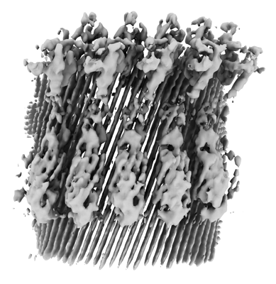 emd_13269.png emd_13269.png | 136.5 KB | ||
| Archive directory |  http://ftp.pdbj.org/pub/emdb/structures/EMD-13269ftp://ftp.pdbj.org/pub/emdb/structures/EMD-13269 http://ftp.pdbj.org/pub/emdb/structures/EMD-13269ftp://ftp.pdbj.org/pub/emdb/structures/EMD-13269 | HTTPS FTP |
-Related structure data
| Related structure data |  7pagMC M: atomic model generated by this map C: citing same article ( |
|---|---|
| Similar structure data |


-Links
| EMDB pages | EMDB (EBI/PDBe) / EMDataResource |
|---|---|
| Related items in Molecule of the Month |


-Map
| File | Download / File: emd_13269.map.gz / Format: CCP4 / Size: 52.7 MB / Type: IMAGE STORED AS FLOATING POINT NUMBER (4 BYTES) | ||||||||||||||||||||||||||||||||||||||||||||||||||||||||||||
|---|---|---|---|---|---|---|---|---|---|---|---|---|---|---|---|---|---|---|---|---|---|---|---|---|---|---|---|---|---|---|---|---|---|---|---|---|---|---|---|---|---|---|---|---|---|---|---|---|---|---|---|---|---|---|---|---|---|---|---|---|---|
| Voxel size | X=Y=Z: 1.34 Å | ||||||||||||||||||||||||||||||||||||||||||||||||||||||||||||
| Density |
| ||||||||||||||||||||||||||||||||||||||||||||||||||||||||||||
| Symmetry | Space group: 1 | ||||||||||||||||||||||||||||||||||||||||||||||||||||||||||||
| Details | EMDB XML:
CCP4 map header:
| ||||||||||||||||||||||||||||||||||||||||||||||||||||||||||||
-Supplemental data
- Sample components
-Entire : Oligomerised mouse Perforin in its inserted state
| Entire | Name: Oligomerised mouse Perforin in its inserted state |
|---|---|
| Components |
|
-Supramolecule #1: Oligomerised mouse Perforin in its inserted state
| Supramolecule | Name: Oligomerised mouse Perforin in its inserted state / type: complex / ID: 1 / Parent: 0 / Macromolecule list: #1 |
|---|---|
| Source (natural) | Organism: Mus musculus (house mouse) |
| Recombinant expression | Organism:   Spodoptera frugiperda (fall armyworm) Spodoptera frugiperda (fall armyworm) |
-Macromolecule #1: Perforin-1
| Macromolecule | Name: Perforin-1 / type: protein_or_peptide / ID: 1 / Number of copies: 1 / Enantiomer: LEVO |
|---|---|
| Source (natural) | Organism: Mus musculus (house mouse) |
| Molecular weight | Theoretical: 60.827051 KDa |
| Recombinant expression | Organism: Spodoptera frugiperda (fall armyworm) |
| Sequence | String: PCYTATRSEC KQKHKFVPGV WMAGEGMDVT TLRRSGSFPV NTQRFLRPDR TCTLCKNSLM RDATQRLPVA ITHWRPHSSH CQRNVAAAK VHSTEGVARE AAANINNDWR VGLDVNPRPE ANMRASVAGS HSKVANFAAE KTYQDQYNFN SDTVECRMYS F RLVQKPPL ...String: PCYTATRSEC KQKHKFVPGV WMAGEGMDVT TLRRSGSFPV NTQRFLRPDR TCTLCKNSLM RDATQRLPVA ITHWRPHSSH CQRNVAAAK VHSTEGVARE AAANINNDWR VGLDVNPRPE ANMRASVAGS HSKVANFAAE KTYQDQYNFN SDTVECRMYS F RLVQKPPL HLDFKKALRA LPRNFNSSTE HAYHRLISSY GTHFITAVDL GGRISVLTAL RTCQLTLNGL TADEVGDCLN VE AQVSIGA QASVSSEYKA CEEKKKQHKM ATSFHQTYRE RHVEVLGGPL DSTHDLLFGN QATPEQFSTW TASLPSNPGL VDY SLEPLH TLLEEQNPKR EALRQAISHY IMSRARWQNC SRPCRSGQHK SSHDSCQCEC QDSKVTNQDC CPRQRGLAHL VVSN FRAEH LWGDYTTATD AYLKVFFGGQ EFRTGVVWNN NNPRWTDKMD FENVLLSTGG PLRVQVWDAD YGWDDDLLGS CDRSP HSGF HEVTCELNHG RVKFSYHAKC LPHLTGGTCL EYAPQGLLGD PPGNRSGAVW HHHHHH |
-Macromolecule #2: CALCIUM ION
| Macromolecule | Name: CALCIUM ION / type: ligand / ID: 2 / Number of copies: 3 / Formula: CA |
|---|---|
| Molecular weight | Theoretical: 40.078 Da |
-Macromolecule #3: 2-acetamido-2-deoxy-beta-D-glucopyranose
| Macromolecule | Name: 2-acetamido-2-deoxy-beta-D-glucopyranose / type: ligand / ID: 3 / Number of copies: 1 / Formula: NAG |
|---|---|
| Molecular weight | Theoretical: 221.208 Da |
| Chemical component information |  ChemComp-NAG: |
-Experimental details
-Structure determination
| Method | cryo EM |
|---|---|
 Processing Processing | single particle reconstruction |
| Aggregation state | particle |
-Sample preparation
| Buffer | pH: 7.5 |
|---|---|
| Grid | Model: UltrAuFoil R2/2 / Material: GOLD / Support film - Material: GRAPHENE OXIDE / Pretreatment - Type: GLOW DISCHARGE |
| Vitrification | Cryogen name: ETHANE |
- Electron microscopy
| Microscope | FEI TITAN KRIOS |
|---|---|
| Electron beam | Acceleration voltage: 300 kV / Electron source: FIELD EMISSION GUN |
| Electron optics | Illumination mode: FLOOD BEAM / Imaging mode: BRIGHT FIELDBright-field microscopy |
| Image recording | Film or detector model: GATAN K3 BIOQUANTUM (6k x 4k) / Average electron dose: 49.6 e/Å2 |
| Experimental equipment |  Model: Titan Krios / Image courtesy: FEI Company |
-Image processing
| Initial angle assignment | Type: MAXIMUM LIKELIHOOD |
|---|---|
| Final angle assignment | Type: MAXIMUM LIKELIHOOD |
| Final reconstruction | Resolution.type: BY AUTHOR / Resolution: 4.0 Å / Resolution method: FSC 0.143 CUT-OFF Details: 229,789 particles were symmetry expanded with C22 symmetry giving the final number of particles 5,055,358 Number images used: 229789 |
| FSC plot (resolution estimation) |  |
