 Movie
Movie Controller
 Structure viewers
 About EMN search


-Search query
-Search result
Showing 1 - 50 of 130 items for (author: gonen, & t.)
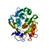
PDB-9cip: 
MicroED structure of the C11 cysteine protease clostripain
Method: electron crystallography / : Ruma YN, Bu G, Hattne J, Gonen T
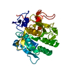
PDB-8vd7:
MicroED structure of SARS-CoV-2 main protease (MPro/3CLPro) with missing cone eliminated by suspended drop
Method: electron crystallography / : Bu G, Gillman C, Danelius E, Hattne J, Nannenga BL, Gonen T

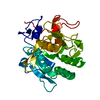
PDB-8w12:
Cryo-EM structure of VP3-VP6 heterohexamer
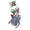
Method: single particle / : Xia X, Sung PY, Martynowycz MW, Gonen T, Roy P, Zhou ZH

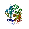
PDB-8w19:
Cryo-EM structure of BTV star-subcore
Method: single particle / : Xia X, Sung PY, Martynowycz MW, Gonen T, Roy P, Zhou ZH

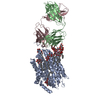
PDB-8w1c:
Cryo-EM structure of BTV pre-subcore
Method: single particle / : Xia X, Sung PY, Martynowycz MW, Gonen T, Roy P, Zhou ZH

PDB-8w1i:
Cryo-EM structure of BTV subcore
Method: single particle / : Xia X, Sung PY, Martynowycz MW, Gonen T, Roy P, Zhou ZH

PDB-8w1o:
Cryo-EM structure of BTV virion
Method: single particle / : Xia X, Sung PY, Martynowycz MW, Gonen T, Roy P, Zhou ZH

PDB-8w1r:
Cryo-EM structure of BTV core
Method: single particle / : Xia X, Sung PY, Martynowycz MW, Gonen T, Roy P, Zhou ZH

PDB-8w1s:
Cryo-EM structure of BTV pre-core
Method: single particle / : Xia X, Sung PY, Martynowycz MW, Gonen T, Roy P, Zhou ZH
PDB-8eum:
MicroED structure of an Aeropyrum pernix protoglobin mutant
Method: electron crystallography / : Danelius E, Gonen T, Unge JT
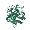
PDB-8sdk:
The MicroED structure of proteinase K crystallized by suspended drop crystallization
Method: electron crystallography / : Gillman C, Nicolas WJ, Martynowycz MW, Gonen T

PDB-8fyo:
MicroED structure of Proteinase K from lamellae milled from multiple plasma sources
Method: electron crystallography / : Martynowycz MW, Shiriaeva A, Clabbers MTB, Nicolas WJ, Weaver SJ, Hattne J, Gonen T
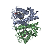
PDB-8d2s:
Zebrafish MFSD2A isoform B in inward open ligand bound conformation
Method: single particle / : Nguyen C, Lei HT, Lai LTF, Gallentino MJ, Mu X, Matthies D, Gonen T
PDB-8d2t:
Zebrafish MFSD2A isoform B in inward open ligand-free conformation
Method: single particle / : Nguyen C, Lei HT, Lai LTF, Gallentino MJ, Mu X, Matthies D, Gonen T

PDB-8d2u:
Zebrafish MFSD2A isoform B in inward open ligand 1A conformation
Method: single particle / : Nguyen C, Lei HT, Lai LTF, Gallentino MJ, Mu X, Matthies D, Gonen T

PDB-8d2v:
Zebrafish MFSD2A isoform B in inward open ligand 1B conformation
Method: single particle / : Nguyen C, Lei HT, Lai LTF, Gallentino MJ, Mu X, Matthies D, Gonen T

PDB-8d2w:
Zebrafish MFSD2A isoform B in inward open ligand 2B conformation
Method: single particle / : Nguyen C, Lei HT, Lai LTF, Gallentino MJ, Mu X, Matthies D, Gonen T

PDB-8d2x:
Zebrafish MFSD2A isoform B in inward open ligand 3C conformation
Method: single particle / : Nguyen C, Lei HT, Lai LTF, Gallentino MJ, Mu X, Matthies D, Gonen T
PDB-8eun:
MicroED structure of an Aeropyrum pernix protoglobin metallo-carbene complex
Method: electron crystallography / : Danelius E, Gonen T, Unge JT

PDB-7uly:
MicroED structure of triclinic lysozyme
Method: electron crystallography / : Clabbers MTB, Martynowycz MW, Hattne J, Gonen T

PDB-8fyn:
MicroED structure of A2A from plasma milled lamellae
Method: electron crystallography / : Martynowycz MW, Shiriaeva A, Clabbers MTB, Nicolas WJ, Weaver SJ, Hattne J, Gonen T

PDB-8fyp:
MicroED structure of Proteinase K from xenon milled lamellae
Method: electron crystallography / : Martynowycz MW, Shiriaeva A, Clabbers MTB, Nicolas WJ, Weaver SJ, Hattne J, Gonen T

PDB-8fyq:
MicroED structure of Proteinase K from argon milled lamellae
Method: electron crystallography / : Martynowycz MW, Shiriaeva A, Clabbers MTB, Nicolas WJ, Weaver SJ, Hattne J, Gonen T

PDB-8fyr:
MicroED structure of Proteinase K from oxygen milled lamellae
Method: electron crystallography / : Martynowycz MW, Shiriaeva A, Clabbers MTB, Nicolas WJ, Weaver SJ, Hattne J, Gonen T

PDB-8fys:
MicroED structure of Proteinase K from nitrogen milled lamellae
Method: electron crystallography / : Martynowycz MW, Shiriaeva A, Clabbers MTB, Nicolas WJ, Weaver SJ, Hattne J, Gonen T
PDB-8e52:
MicroED structure of proteinase K recorded on K2
Method: electron crystallography / : Clabbers MTB, Martynowycz MW, Hattne J, Nannenga BL, Gonen T

PDB-8e53:
MicroED structure of proteinase K recorded on K3
Method: electron crystallography / : Clabbers MTB, Martynowycz MW, Hattne J, Nannenga BL, Gonen T

PDB-8e54:
MicroED structure of triclinic lysozyme recorded on K3
Method: electron crystallography / : Clabbers MTB, Martynowycz MW, Hattne J, Nannenga BL, Gonen T
PDB-7svy:
MicroED structure of proteinase K from a 130 nm thick lamella measured at 120 kV
Method: electron crystallography / : Martynowycz MW, Clabbers MTB, Unge J, Hattne J, Gonen T
PDB-7svz:
MicroED structure of proteinase K from a 200 nm thick lamella measured at 120 kV
Method: electron crystallography / : Martynowycz MW, Clabbers MTB, Unge J, Hattne J, Gonen T
PDB-7sw0:
MicroED structure of proteinase K from a 325 nm thick lamella measured at 120 kV
Method: electron crystallography / : Martynowycz MW, Clabbers MTB, Unge J, Hattne J, Gonen T
PDB-7sw1:
MicroED structure of proteinase K from a 115 nm thick lamella measured at 200 kV
Method: electron crystallography / : Martynowycz MW, Clabbers MTB, Unge J, Hattne J, Gonen T
PDB-7sw2:
MicroED structure of proteinase K from a 130 nm thick lamella measured at 200 kV
Method: electron crystallography / : Martynowycz MW, Clabbers MTB, Unge J, Hattne J, Gonen T

PDB-7sw3:
MicroED structure of proteinase K from a 95 nm thick lamella measured at 200 kV
Method: electron crystallography / : Martynowycz MW, Clabbers MTB, Unge J, Hattne J, Gonen T

PDB-7sw4:
MicroED structure of proteinase K from a 540 nm thick lamella measured at 200 kV
Method: electron crystallography / : Martynowycz MW, Clabbers MTB, Unge J, Hattne J, Gonen T

PDB-7sw5:
MicroED structure of proteinase K from a 460 nm thick lamella measured at 200 kV
Method: electron crystallography / : Martynowycz MW, Clabbers MTB, Unge J, Hattne J, Gonen T
PDB-7sw6:
MicroED structure of proteinase K from a 260 nm thick lamella measured at 200 kV
Method: electron crystallography / : Martynowycz MW, Clabbers MTB, Unge J, Hattne J, Gonen T
PDB-7sw7:
MicroED structure of proteinase K from a 530 nm thick lamella measured at 200 kV
Method: electron crystallography / : Martynowycz MW, Clabbers MTB, Unge J, Hattne J, Gonen T
PDB-7sw8:
MicroED structure of proteinase K from a 150 nm thick lamella measured at 300 kV
Method: electron crystallography / : Martynowycz MW, Clabbers MTB, Unge J, Hattne J, Gonen T

PDB-7sw9:
MicroED structure of proteinase K from a 170 nm thick lamella measured at 300 kV
Method: electron crystallography / : Martynowycz MW, Clabbers MTB, Unge J, Hattne J, Gonen T
PDB-7swa:
MicroED structure of proteinase K from a 320 nm thick lamella measured at 300 kV
Method: electron crystallography / : Martynowycz MW, Clabbers MTB, Unge J, Hattne J, Gonen T

PDB-7swb:
MicroED structure of proteinase K from a 360 nm thick lamella measured at 300 kV
Method: electron crystallography / : Martynowycz MW, Clabbers MTB, Unge J, Hattne J, Gonen T
PDB-7swc:
MicroED structure of proteinase K from a 550 nm thick lamella measured at 300 kV
Method: electron crystallography / : Martynowycz MW, Clabbers MTB, Unge J, Hattne J, Gonen T
PDB-7skw:
Ab initio structure of triclinic lysozyme from electron-counted MicroED data
Method: electron crystallography / : Martynowycz MW, Clabbers MTB, Hattne J, Gonen T

PDB-7skx:
Ab initio structure of proteinase K from electron-counted MicroED data
Method: electron crystallography / : Martynowycz MW, Clabbers MTB, Hattne J, Gonen T
PDB-7ute:
MicroED structure of Aeropyrum pernix protoglobin mutant
Method: electron crystallography / : Danelius E, Gonen T, Unge JT
PDB-7mrp:
MicroED structure of lysozyme from milled crystals at 1.75A
Method: electron crystallography / : Martynowycz MW, Gonen T

PDB-7rm5:
MicroED structure of the human adenosine receptor at 2.8A
Method: electron crystallography / : Martynowycz MW, Shiriaeva A, Ge X, Hattne J, Nannenga BL, Cherezov V, Gonen T

PDB-7kuh:
MicroED structure of mVDAC
Method: electron crystallography / : Martynowycz MW, Khan F, Hattne J, Abramson J, Gonen T

PDB-7jsy:
Proteinase K soaked with I3C determined by MicroED from a single milled microcrystal
Method: electron crystallography / : Martynowycz MW, Gonen T
Pages:
 wwPDB to switch to version 3 of the EMDB data model
