 Movie
Movie Controller

[English] 日本語
 Yorodumi
- PDB-7skw: Ab initio structure of triclinic lysozyme from electron-counted M... -
+ Open data

- Basic information
| Entry | Database: PDB / ID: 7skw | |||||||||
|---|---|---|---|---|---|---|---|---|---|---|
| Title | Ab initio structure of triclinic lysozyme from electron-counted MicroED data | |||||||||
 Components Components | Lysozyme C | |||||||||
 Keywords Keywords |  HYDROLASE HYDROLASE | |||||||||
| Function / homology |  Function and homology information Function and homology informationLactose synthesis / Antimicrobial peptides / Neutrophil degranulation / beta-N-acetylglucosaminidase activity / cell wall macromolecule catabolic process / lysozyme / lysozyme activity / killing of cells of another organism / defense response to Gram-negative bacterium / defense response to Gram-positive bacterium ...Lactose synthesis / Antimicrobial peptides / Neutrophil degranulation / beta-N-acetylglucosaminidase activity / cell wall macromolecule catabolic process / lysozyme / lysozyme activity / killing of cells of another organism / defense response to Gram-negative bacterium / defense response to Gram-positive bacterium / defense response to bacterium / endoplasmic reticulum / extracellular space / identical protein binding / cytoplasmSimilarity search - Function | |||||||||
| Biological species |  Gallus gallus (chicken) Gallus gallus (chicken) | |||||||||
| Method | ELECTRON CRYSTALLOGRAPHY / electron crystallography / cryo EM / Resolution: 0.87 Å | |||||||||
 Authors Authors | Martynowycz, M.W. / Clabbers, M.T.B. / Hattne, J. / Gonen, T. | |||||||||
| Funding support |  United States, 2items United States, 2items
| |||||||||
 Citation Citation | Journal: Nat Methods / Year: 2022 Title: Ab initio phasing macromolecular structures using electron-counted MicroED data. Authors: Michael W Martynowycz / Max T B Clabbers / Johan Hattne / Tamir Gonen / Abstract: Structures of two globular proteins were determined ab initio using microcrystal electron diffraction (MicroED) data that were collected on a direct electron detector in counting mode. Microcrystals ...Structures of two globular proteins were determined ab initio using microcrystal electron diffraction (MicroED) data that were collected on a direct electron detector in counting mode. Microcrystals were identified using a scanning electron microscope (SEM) and thinned with a focused ion beam (FIB) to produce crystalline lamellae of ideal thickness. Continuous-rotation data were collected using an ultra-low exposure rate to enable electron counting in diffraction. For the first sample, triclinic lysozyme extending to a resolution of 0.87 Å, an ideal helical fragment of only three alanine residues provided initial phases. These phases were improved using density modification, allowing the entire atomic structure to be built automatically. A similar approach was successful on a second macromolecular sample, proteinase K, which is much larger and diffracted to a resolution of 1.5 Å. These results demonstrate that macromolecules can be determined to sub-ångström resolution by MicroED and that ab initio phasing can be successfully applied to counting data. | |||||||||
| History |
|
- Structure visualization
| Structure viewer | Molecule: MolmilJmol/JSmol |
|---|
- Downloads & links
-Download
| PDBx/mmCIF format | 7skw.cif.gz | 104.9 KB | Display | PDBx/mmCIF format |
|---|---|---|---|---|
| PDB format | pdb7skw.ent.gz | 79.3 KB | Display | PDB format |
| PDBx/mmJSON format | 7skw.json.gz | Tree view | PDBx/mmJSON format | |
| Others |  Other downloads Other downloads |
-Validation report
| Arichive directory | https://data.pdbj.org/pub/pdb/validation_reports/sk/7skwftp://data.pdbj.org/pub/pdb/validation_reports/sk/7skw | HTTPS FTP |
|---|
-Related structure data
| Related structure data |  25184MC  7skxC M: map data used to model this data C: citing same article ( |
|---|---|
| Similar structure data |

-Links
 PDBj
PDBj


- Assembly
| Deposited unit | 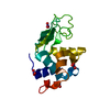
| ||||||||
|---|---|---|---|---|---|---|---|---|---|
| 1 |
| ||||||||
| Unit cell |
|
-Components
| #1: Protein | Mass: 14331.160 Da / Num. of mol.: 1 / Source method: isolated from a natural source / Source: (natural) Gallus gallus (chicken) / References: UniProt: P00698, lysozyme | ||||
|---|---|---|---|---|---|
| #2: Chemical | ChemComp-NO3 / Nitrate  Mass: 62.005 Da / Num. of mol.: 4 / Source method: obtained synthetically / Formula: NO3 Mass: 62.005 Da / Num. of mol.: 4 / Source method: obtained synthetically / Formula: NO3#3: Water | ChemComp-HOH / | Water Mass: 18.015 Da / Num. of mol.: 156 / Source method: isolated from a natural source / Formula: H2O Mass: 18.015 Da / Num. of mol.: 156 / Source method: isolated from a natural source / Formula: H2OHas ligand of interest | N | |
-Experimental details
-Experiment
| Experiment | Method: ELECTRON CRYSTALLOGRAPHY |
|---|---|
| EM experiment | Aggregation state: 3D ARRAY / 3D reconstruction method: electron crystallography |
- Sample preparation
| Component | Name: Lysozyme / Type: COMPLEX / Entity ID: #1 / Source: NATURAL |
|---|---|
| Molecular weight | Value: 0.0144 MDa / Experimental value: NO |
| Source (natural) | Organism: Gallus gallus (chicken) |
| Buffer solution | pH: 4.7 |
| Specimen | Conc.: 5 mg/ml / Embedding applied: NO / Shadowing applied: NO / Staining applied: NO / Vitrification applied: YES / Details: Milled microcrystals |
| Specimen support | Grid material: COPPER / Grid mesh size: 200 divisions/in. / Grid type: Quantifoil R2/2 |
| Vitrification | Instrument: LEICA PLUNGER / Cryogen name: ETHANE / Humidity: 95 % / Chamber temperature: 277 K |
-Data collection
| Experimental equipment |  Model: Titan Krios / Image courtesy: FEI Company |
|---|---|
| Microscopy | Model: FEI TITAN KRIOS |
| Electron gun | Electron source: FIELD EMISSION GUN / Accelerating voltage: 300 kV / Illumination mode: FLOOD BEAM |
| Electron lens | Mode: DIFFRACTION / Cs: 2.7 mm / C2 aperture diameter: 50 µm / Alignment procedure: BASIC |
| Specimen holder | Cryogen: NITROGEN / Specimen holder model: FEI TITAN KRIOS AUTOGRID HOLDER / Temperature (max): 90 K / Temperature (min): 77 K |
| Image recording | Average exposure time: 0.5 sec. / Electron dose: 0.001 e/Å2 / Film or detector model: FEI FALCON IV (4k x 4k) / Num. of diffraction images: 840 / Num. of grids imaged: 1 / Num. of real images: 1 Details: 0.15 degrees per second, 0.5 second readout, 30 to -30 degrees |
| Image scans | Sampling size: 28 µm / Width: 2048 / Height: 2048 |
| EM diffraction | Camera length: 1202 mm / Tilt angle list: -30,30 |
| EM diffraction shell | Resolution: 0.87→0.9 Å / Fourier space coverage: 37.64 % / Multiplicity: 2.1 / Num. of structure factors: 2783 / Phase residual: 30 ° |
| EM diffraction stats | Fourier space coverage: 87.58 % / High resolution: 0.87 Å / Num. of intensities measured: 569407 / Num. of structure factors: 64974 / Phase error: 30 ° / Phase error rejection criteria: None / Rmerge: 0.236 / Rsym: 0.073 |
- Processing
| Software | Name: REFMAC / Version: 5.8.0267 / Classification: refinement / Contact author: Garib N. Murshudov / Contact author email: garib[at]mrc-lmb.cam.ac.uk / Date: 2020-24-08 Description: (un)restrained refinement or idealisation of macromolecular structures | ||||||||||||||||||||||||||||||||||||||||||||||||||||||||||||||||||||||||||||||||||||||||||||||||||||||||||
|---|---|---|---|---|---|---|---|---|---|---|---|---|---|---|---|---|---|---|---|---|---|---|---|---|---|---|---|---|---|---|---|---|---|---|---|---|---|---|---|---|---|---|---|---|---|---|---|---|---|---|---|---|---|---|---|---|---|---|---|---|---|---|---|---|---|---|---|---|---|---|---|---|---|---|---|---|---|---|---|---|---|---|---|---|---|---|---|---|---|---|---|---|---|---|---|---|---|---|---|---|---|---|---|---|---|---|---|
| EM software |
| ||||||||||||||||||||||||||||||||||||||||||||||||||||||||||||||||||||||||||||||||||||||||||||||||||||||||||
| Image processing | Details: Binned by 2 | ||||||||||||||||||||||||||||||||||||||||||||||||||||||||||||||||||||||||||||||||||||||||||||||||||||||||||
| EM 3D crystal entity | ∠α: 88.319 ° / ∠β: 109.095 ° / ∠γ: 112.075 ° / A: 26.42 Å / B: 30.72 Å / C: 33.01 Å / Space group name: P1 / Space group num: 1 | ||||||||||||||||||||||||||||||||||||||||||||||||||||||||||||||||||||||||||||||||||||||||||||||||||||||||||
| CTF correction | Type: NONE | ||||||||||||||||||||||||||||||||||||||||||||||||||||||||||||||||||||||||||||||||||||||||||||||||||||||||||
| 3D reconstruction | Resolution method: DIFFRACTION PATTERN/LAYERLINES / Symmetry type: 3D CRYSTAL | ||||||||||||||||||||||||||||||||||||||||||||||||||||||||||||||||||||||||||||||||||||||||||||||||||||||||||
| Atomic model building | B value: 12.188 / Protocol: AB INITIO MODEL / Space: RECIPROCAL / Target criteria: Maximum likelihood | ||||||||||||||||||||||||||||||||||||||||||||||||||||||||||||||||||||||||||||||||||||||||||||||||||||||||||
| Refinement | Resolution: 0.87→19.876 Å / Cor.coef. Fo:Fc: 0.951 / Cor.coef. Fo:Fc free: 0.951 / SU B: 1.263 / SU ML: 0.03 / Cross valid method: FREE R-VALUE / ESU R: 0.028 / ESU R Free: 0.028 Details: Hydrogens have been added in their riding positions
| ||||||||||||||||||||||||||||||||||||||||||||||||||||||||||||||||||||||||||||||||||||||||||||||||||||||||||
| Solvent computation | Ion probe radii: 0.8 Å / Shrinkage radii: 0.8 Å / VDW probe radii: 1.2 Å / Solvent model: MASK BULK SOLVENT | ||||||||||||||||||||||||||||||||||||||||||||||||||||||||||||||||||||||||||||||||||||||||||||||||||||||||||
| Displacement parameters | Biso mean: 12.188 Å2
| ||||||||||||||||||||||||||||||||||||||||||||||||||||||||||||||||||||||||||||||||||||||||||||||||||||||||||
| Refine LS restraints |
|