 Movie
Movie Controller

[English] 日本語
 Yorodumi
- PDB-7sw6: MicroED structure of proteinase K from a 260 nm thick lamella mea... -
+ Open data

- Basic information
| Entry | Database: PDB / ID: 7sw6 | |||||||||
|---|---|---|---|---|---|---|---|---|---|---|
| Title | MicroED structure of proteinase K from a 260 nm thick lamella measured at 200 kV | |||||||||
 Components Components | Proteinase K | |||||||||
 Keywords Keywords | HYDROLASE / Serine protease | |||||||||
| Function / homology |  Function and homology information Function and homology informationpeptidase K / serine-type endopeptidase activity / proteolysis / extracellular region / metal ion bindingSimilarity search - Function | |||||||||
| Biological species |  Parengyodontium album (fungus) Parengyodontium album (fungus) | |||||||||
| Method | ELECTRON CRYSTALLOGRAPHY / electron crystallography / MOLECULAR REPLACEMENT / cryo EM / Resolution: 1.95 Å | |||||||||
 Authors Authors | Martynowycz, M.W. / Clabbers, M.T.B. / Unge, J. / Hattne, J. / Gonen, T. | |||||||||
| Funding support |  United States, 2items United States, 2items
| |||||||||
 Citation Citation | Journal: Proc Natl Acad Sci U S A / Year: 2021 Title: Benchmarking the ideal sample thickness in cryo-EM. Authors: Michael W Martynowycz / Max T B Clabbers / Johan Unge / Johan Hattne / Tamir Gonen / Abstract: The relationship between sample thickness and quality of data obtained is investigated by microcrystal electron diffraction (MicroED). Several electron microscopy (EM) grids containing proteinase K ...The relationship between sample thickness and quality of data obtained is investigated by microcrystal electron diffraction (MicroED). Several electron microscopy (EM) grids containing proteinase K microcrystals of similar sizes from the same crystallization batch were prepared. Each grid was transferred into a focused ion beam and a scanning electron microscope in which the crystals were then systematically thinned into lamellae between 95- and 1,650-nm thick. MicroED data were collected at either 120-, 200-, or 300-kV accelerating voltages. Lamellae thicknesses were expressed in multiples of the corresponding inelastic mean free path to allow the results from different acceleration voltages to be compared. The quality of the data and subsequently determined structures were assessed using standard crystallographic measures. Structures were reliably determined with similar quality from crystalline lamellae up to twice the inelastic mean free path. Lower resolution diffraction was observed at three times the mean free path for all three accelerating voltages, but the data quality was insufficient to yield structures. Finally, no coherent diffraction was observed from lamellae thicker than four times the calculated inelastic mean free path. This study benchmarks the ideal specimen thickness with implications for all cryo-EM methods. | |||||||||
| History |
|
- Structure visualization
| Structure viewer | Molecule: MolmilJmol/JSmol |
|---|
- Downloads & links
-Download
| PDBx/mmCIF format | 7sw6.cif.gz | 83.7 KB | Display | PDBx/mmCIF format |
|---|---|---|---|---|
| PDB format | pdb7sw6.ent.gz | 47.5 KB | Display | PDB format |
| PDBx/mmJSON format | 7sw6.json.gz | Tree view | PDBx/mmJSON format | |
| Others |  Other downloads Other downloads |
-Validation report
| Arichive directory | https://data.pdbj.org/pub/pdb/validation_reports/sw/7sw6ftp://data.pdbj.org/pub/pdb/validation_reports/sw/7sw6 | HTTPS FTP |
|---|
-Related structure data
| Related structure data |  25464MC 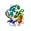 7svyC 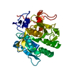 7svzC 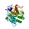 7sw0C 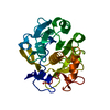 7sw1C 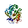 7sw2C  7sw3C  7sw4C  7sw5C 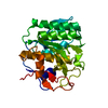 7sw7C 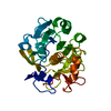 7sw8C  7sw9C 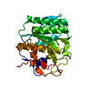 7swaC  7swbC 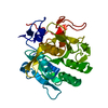 7swcC 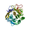 6cl7S |
|---|---|
| Similar structure data |


-Links
 PDBj
PDBj

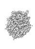
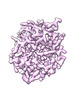
- Assembly
| Deposited unit | 
| ||||||||||||
|---|---|---|---|---|---|---|---|---|---|---|---|---|---|
| 1 |
| ||||||||||||
| Unit cell |
| ||||||||||||
| Components on special symmetry positions |
|
-Components
| #1: Protein | / Endopeptidase K / Tritirachium alkaline proteinase Mass: 28930.783 Da / Num. of mol.: 1 / Source method: isolated from a natural source / Source: (natural) Parengyodontium album (fungus) / References: UniProt: P06873, peptidase K |
|---|---|
| #2: Water | ChemComp-HOH / Water Mass: 18.015 Da / Num. of mol.: 127 / Source method: isolated from a natural source / Formula: H2O Mass: 18.015 Da / Num. of mol.: 127 / Source method: isolated from a natural source / Formula: H2O |
-Experimental details
-Experiment
| Experiment | Method: ELECTRON CRYSTALLOGRAPHY |
|---|---|
| EM experiment | Aggregation state: 3D ARRAY / 3D reconstruction method: electron crystallography |
- Sample preparation
| Component | Name: Proteinase K / Type: COMPLEX / Details: Serine protease / Entity ID: #1 / Source: NATURAL |
|---|---|
| Molecular weight | Value: 0.0289 MDa / Experimental value: NO |
| Source (natural) | Organism: Parengyodontium album (fungus) |
| Buffer solution | pH: 7.5 |
| Specimen | Conc.: 5 mg/ml / Embedding applied: NO / Shadowing applied: NO / Staining applied: NO / Vitrification applied: YES / Details: Microcrystals |
| Specimen support | Grid material: COPPER / Grid mesh size: 200 divisions/in. / Grid type: Quantifoil R2/2 |
| Vitrification | Instrument: LEICA PLUNGER / Cryogen name: ETHANE / Humidity: 95 % / Chamber temperature: 277 K |
-Data collection
| Experimental equipment |  Model: Talos Arctica / Image courtesy: FEI Company |
|---|---|
| Microscopy | Model: FEI TALOS ARCTICA |
| Electron gun | Electron source: FIELD EMISSION GUN / Accelerating voltage: 200 kV / Illumination mode: FLOOD BEAM |
| Electron lens | Mode: DIFFRACTION / C2 aperture diameter: 100 µm / Alignment procedure: BASIC |
| Specimen holder | Cryogen: NITROGEN / Specimen holder model: FEI TITAN KRIOS AUTOGRID HOLDER / Temperature (max): 90 K / Temperature (min): 77 K |
| Image recording | Average exposure time: 1 sec. / Electron dose: 0.01 e/Å2 / Film or detector model: FEI CETA (4k x 4k) / Num. of diffraction images: 120 / Num. of grids imaged: 1 / Num. of real images: 1 Details: 0.5 degrees per second, 1 second readout, 30 to -30 degrees. |
| Image scans | Sampling size: 28 µm / Width: 2048 / Height: 2048 |
| EM diffraction | Camera length: 1863 mm |
| EM diffraction shell | Resolution: 1.95→2.07 Å / Fourier space coverage: 92 % / Multiplicity: 5 / Num. of structure factors: 2561 / Phase residual: 29 ° |
| EM diffraction stats | Fourier space coverage: 91 % / High resolution: 1.95 Å / Num. of intensities measured: 88107 / Num. of structure factors: 16540 / Phase error: 22 ° / Phase error rejection criteria: None / Rmerge: 28 / Rsym: 13 |
| Detector | Date: Nov 14, 2020 |
| Reflection | Resolution: 1.95→40.91 Å / Num. obs: 88107 / % possible obs: 91.1 % / Redundancy: 5.3 % / Biso Wilson estimate: 22.29 Å2 / CC1/2: 0.976 / Rmerge(I) obs: 0.277 / Rpim(I) all: 0.133 / Rrim(I) all: 0.309 / Net I/σ(I): 4.25 |
| Reflection shell | Resolution: 1.95→2 Å / Num. unique obs: 6241 / CC1/2: 0.411 / Rrim(I) all: 1.514 / Net I/σ(I) obs: 0.9 / % possible all: 92.7 |
- Processing
| Software |
| |||||||||||||||||||||||||||||||||||||||||||||||||
|---|---|---|---|---|---|---|---|---|---|---|---|---|---|---|---|---|---|---|---|---|---|---|---|---|---|---|---|---|---|---|---|---|---|---|---|---|---|---|---|---|---|---|---|---|---|---|---|---|---|---|
| EM software |
| |||||||||||||||||||||||||||||||||||||||||||||||||
| Image processing | Details: Binned by 2. | |||||||||||||||||||||||||||||||||||||||||||||||||
| EM 3D crystal entity | ∠α: 90 ° / ∠β: 90 ° / ∠γ: 90 ° / A: 67.91 Å / B: 67.91 Å / C: 102.52 Å / Space group name: P43212 / Space group num: 96 | |||||||||||||||||||||||||||||||||||||||||||||||||
| CTF correction | Type: NONE | |||||||||||||||||||||||||||||||||||||||||||||||||
| 3D reconstruction | Resolution method: DIFFRACTION PATTERN/LAYERLINES / Symmetry type: 3D CRYSTAL | |||||||||||||||||||||||||||||||||||||||||||||||||
| Atomic model building | B value: 20 / Protocol: RIGID BODY FIT / Space: RECIPROCAL / Target criteria: Maximum likelihood | |||||||||||||||||||||||||||||||||||||||||||||||||
| Atomic model building | PDB-ID: 6CL7 Pdb chain-ID: A / Accession code: 6CL7 / Pdb chain residue range: 106-384 / Source name: PDB / Type: experimental model | |||||||||||||||||||||||||||||||||||||||||||||||||
| Refinement | Method to determine structure: MOLECULAR REPLACEMENT Starting model: 6CL7 Resolution: 1.95→30.53 Å / SU ML: 0.2566 / Cross valid method: FREE R-VALUE / σ(F): 1.36 / Phase error: 22.3984 Stereochemistry target values: GeoStd + Monomer Library + CDL v1.2
| |||||||||||||||||||||||||||||||||||||||||||||||||
| Solvent computation | Shrinkage radii: 0.9 Å / VDW probe radii: 1.11 Å / Solvent model: FLAT BULK SOLVENT MODEL | |||||||||||||||||||||||||||||||||||||||||||||||||
| Displacement parameters | Biso mean: 20.03 Å2 | |||||||||||||||||||||||||||||||||||||||||||||||||
| Refinement step | Cycle: LAST / Resolution: 1.95→30.53 Å
| |||||||||||||||||||||||||||||||||||||||||||||||||
| Refine LS restraints |
| |||||||||||||||||||||||||||||||||||||||||||||||||
| LS refinement shell |
|
