 Movie
Movie Controller
 Structure viewers
 About EMN search


-Search query
-Search result
Showing all 42 items for (author: e. & v. & orlova)

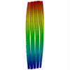
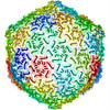
PDB-7ywh: 
Six DNA Helix Bundle nanopore - State 1
Method: single particle / : Javed A, Ahmad K, Lanphere C, Coveney P, Howorka S, Orlova EV
PDB-7ywi:
Six DNA duplex bundle nanopore - State 2
Method: single particle / : Javed A, Ahmad K, Lanphere C, Coveney P, Howorka S, Orlova EV

PDB-7ywl:
Six DNA Helix Bundle nanopore - State 3
Method: single particle / : Javed A, Ahmad K, Lanphere C, Coveney P, Howorka S, Orlova EV

PDB-7ywn:
Six DNA Helix Bundle nanopore - State 4
Method: single particle / : Javed A, Ahmad K, Lanphere C, Orlova EV, Coveney P, Howorka S

PDB-7ywo:
Six DNA Helix Bundle nanopore - State 5
Method: single particle / : Javed A, Ahmad K, Lanphere C, Coveney P, Howorka S, Orlova EV
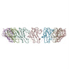
PDB-7z4w:
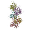
gp6/gp15/gp16 connector complex of bacteriophage SPP1
Method: single particle / : Orlov I, Roche S, Tavares P, Orlova EV

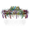
PDB-7pv2:
GA1 bacteriophage portal protein
Method: single particle / : Javed A, Villanueva H, Orlova EV, Savva R

PDB-7pv4:
PhiCPV4 bacteriophage Portal Protein
Method: single particle / : Javed A, Villanueva H, Orlova EV, Savva R

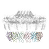
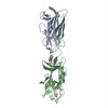
PDB-7apd:
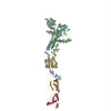
Bovine Papillomavirus E1 DNA helicase-replication fork complex
Method: single particle / : Javed A, Major B, Stead J, Sanders CM, Orlova EV
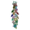
PDB-6hqe:
Cryo-EM of self-assembly peptide filament LRV_M3delta1
Method: helical / : Osinski T, Wang F, Hughes SA, Kreutzberger MAB, Conticello VP, Egelman EH

PDB-6mk1:
Cryo-EM of self-assembly peptide filament HEAT_R1
Method: helical / : Wang F, Hughes SA, Orlova A, Conticello VP, Egelman EH
PDB-5a79:
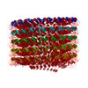
Novel inter-subunit contacts in Barley Stripe Mosaic Virus revealed by cryo-EM
Method: single particle / : Clare DK, Pechnikova E, Skurat E, Makarov V, Sokolova OS, Solovyev AG, V Orlova E
PDB-5a7a:
Novel inter-subunit contacts in Barley Stripe Mosaic Virus revealed by cryo-EM
Method: single particle / : Clare DK, Pechnikova E, Skurat E, Makarov V, Sokolova OS, Solovyev AG, V Orlova E

PDB-5a9k:
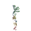
Structural basis for DNA strand separation by a hexameric replicative helicase
Method: single particle / : Chaban Y, Stead JA, Ryzhenkova K, Whelan F, Lamber K, Antson F, Sanders CM, Orlova EV
PDB-5a20:
Structure of bacteriophage SPP1 head-to-tail interface filled with DNA and tape measure protein
Method: single particle / : Chaban Y, Lurz R, Brasiles S, Cornilleau C, Karreman M, Zinn-Justin S, Tavares P, Orlova EV
PDB-5a21:
Structure of bacteriophage SPP1 head-to-tail interface without DNA and tape measure protein
Method: single particle / : Chaban Y, Lurz R, Brasiles S, Cornilleau C, Karreman M, Zinn-Justin S, Tavares P, Orlova EV
PDB-3j8j:
Tilted state of actin, T1
Method: helical / : Galkin VE, Orlova A, Vos MR, Schroder GF, Egelman EH
PDB-3j8k:
Tilted state of actin, T2
Method: helical / : Galkin VE, Orlova A, Vos MR, Schroder GF, Egelman EH
PDB-2m5k:
Atomic-resolution structure of a doublet cross-beta amyloid fibril
Method: single particle / : Fitzpatrick AWP, Debelouchina GT, Bayro MJ, Clare DK, Caporini MA, Bajaj VS, Jaroniec CP, Wang L, Ladizhansky V, Muller S, MacPhee CE, Waudby CA, Mott HR, de Simone A, Knowles TPJ, Saibil HR, Vendruscolo M, Orlova EV, Griffin RG, Dobson CM
PDB-2m5m:
Atomic-resolution structure of a triplet cross-beta amyloid fibril
Method: single particle / : Fitzpatrick AWP, Debelouchina GT, Bayro MJ, Clare DK, Caporini MA, Bajaj VS, Jaroniec CP, Wang L, Ladizhansky V, Muller S, MacPhee CE, Waudby CA, Mott HR, de Simone A, Knowles TPJ, Saibil HR, Vendruscolo M, Orlova EV, Griffin RG, Dobson CM
PDB-3zpk:
Atomic-resolution structure of a quadruplet cross-beta amyloid fibril.
Method: single particle / : Fitzpatrick AWP, Debelouchina GT, Bayro MJ, Clare DK, Caporini MA, Bajaj VS, Jaroniec CP, Wang L, Ladizhansky V, Muller SA, MacPhee CE, Waudby CA, Mott HR, de Simone A, Knowles TPJ, Saibil HR, Vendruscolo M, Orlova EV, Griffin RG, Dobson CM
PDB-2ypw:
Atomic model for the N-terminus of TraO fitted in the full-length structure of the bacterial pKM101 type IV secretion system core complex
Method: single particle / : Rivera-Calzada A, Fronzes R, Savva CG, Chandran V, Lian PW, Laeremans T, Pardon E, Steyaert J, Remaut H, Waksman G, Orlova EV
PDB-3zbi:
Fitting result in the O-layer of the subnanometer structure of the bacterial pKM101 type IV secretion system core complex digested with elastase
Method: single particle / : Rivera-Calzada A, Fronzes R, Savva CG, Chandran V, Lian PW, Laeremans T, Pardon E, Steyaert J, Remaut H, Waksman G, Orlova EV
PDB-3zbj:
Fitting results in the I-layer of the subnanometer structure of the bacterial pKM101 type IV secretion system core complex digested with elastase
Method: single particle / : Rivera-Calzada A, Fronzes R, Savva CG, Chandran V, Lian PW, Laeremans T, Pardon E, Steyaert J, Remaut H, Waksman G, Orlova EV
PDB-4an5:
Capsid structure and its Stability at the Late Stages of Bacteriophage SPP1 Assembly
Method: single particle / : White HE, Sherman MB, Brasiles S, Jacquet E, Seavers P, Tavares P, Orlova EV
PDB-3j0s:
Remodeling of actin filaments by ADF cofilin proteins
Method: helical / : Galkin VE, Orlova A, Kudryashov DS, Solodukhin A, Reisler E, Schroeder GF, Egelman EH

PDB-2xea:
4.6 ANGSTROM CRYO-EM RECONSTRUCTION OF TOBACCO MOSAIC VIRUS FROM IMAGES RECORDED AT 300 KEV ON A 4KX4K CCD CAMERA
Method: helical / : Clare DK, Orlova EV

PDB-3lue:
Model of alpha-actinin CH1 bound to F-actin
Method: helical / : Galkin VE, Orlova A, Salmazo A, Djinovic-Carugo K, Egelman EH

PDB-3iku:
Structural model of ParM filament in closed state from cryo-EM
Method: helical / : Galkin VE, Orlova A, Rivera C, Mullins RD, Egelman EH

PDB-3iky:
Structural model of ParM filament in the open state by cryo-EM
Method: helical / : Galkin VE, Orlova A, Rivera C, Mullins RD, Egelman EH
PDB-3cre:
Electron Microscopy model of the Saf Pilus- Type A
Method: helical / : Salih O, Remaut H, Waksman G, Orlova EV
PDB-3crf:
Electron Microscopy model of the Saf Pilus- Type B
Method: helical / : Salih O, Remaut H, Waksman G, Orlova EV

PDB-3byh:
Model of actin-fimbrin ABD2 complex
Method: helical / : Galkin VE, Orlova A, Cherepanova O, Lebart MC, Egelman EH
PDB-2qu4:
Model for Bacterial ParM Filament
Method: helical / : Orlova A, Garner EC, Galkin VE, Heuser J, Mullins RD, Egelman EH

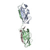
PDB-2j9i:
Lengsin is a survivor of an ancient family of class I glutamine synthetases in eukaryotes that has undergone evolutionary re- engineering for a tissue-specific role in the vertebrate eye lens.
Method: single particle / : Wyatt K, White HE, Wang L, Bateman OA, Slingsby C, Orlova EV, Wistow G
PDB-2h50:
Multiple distinct assemblies reveal conformational flexibility in the small heat shock protein Hsp26
Method: single particle / : White HE, Orlova EV, Chen S, Wang L, Ignatiou A, Gowen B, Stromer T, Franzmann TM, Haslbeck M, Buchner J, Saibil HR
PDB-2h53:
Multiple distinct assemblies reveal conformational flexibility in the small heat shock protein Hsp26
Method: single particle / : White HE, Orlova EV, Chen S, Wang L, Ignatiou A, Gowen B, Stromer T, Franzmann TM, Haslbeck M, Buchner J, Saibil HR

PDB-2byu:
Negative stain EM reconstruction of M.tuberculosis Acr1(Hsp 16.3) fitted with wheat sHSP dimer
Method: single particle / : Kennaway CK, Benesch JLP, Gohlke U, Wang L, Robinson CV, Orlova EV, Saibil HR, Keep NH
PDB-2bk1:
The pore structure of pneumolysin, obtained by fitting the alpha carbon trace of perfringolysin O into a cryo-EM map
Method: single particle / : Tilley SJ, Orlova EV, Gilbert RJC, Andrew PW, Saibil HR

PDB-2bk2:
The prepore structure of pneumolysin, obtained by fitting the alpha carbon trace of perfringolysin O into a cryo-EM map
Method: single particle / : Tilley SJ, Orlova EV, Gilbert RJC, Andrew PW, Saibil HR
PDB-1ml5:
Structure of the E. coli ribosomal termination complex with release factor 2
Method: single particle / : Klaholz BP, Pape T, Zavialov AV, Myasnikov AG, Orlova EV, Vestergaard B, Ehrenberg M, van Heel M
 wwPDB to switch to version 3 of the EMDB data model
