 Movie
Movie Controller


+ Open data

- Basic information
| Entry | Database: PDB / ID: 3iky | ||||||
|---|---|---|---|---|---|---|---|
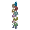
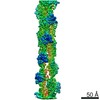
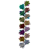
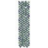
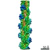
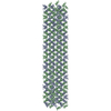
| Title | Structural model of ParM filament in the open state by cryo-EM | ||||||
 Components Components | Plasmid segregation protein parM | ||||||
 Keywords Keywords | STRUCTURAL PROTEIN / polymorphic protein polymers / Plasmid / Plasmid partition | ||||||
| Function / homology | Plasmid segregation protein ParM/StbA / : / Plasmid segregation protein ParM, N-terminal / Plasmid segregation protein ParM, C-terminal / ParM-like / plasmid partitioning / ATPase, nucleotide binding domain / identical protein binding / Plasmid segregation protein ParM Function and homology information Function and homology information | ||||||
| Biological species |  | ||||||
| Method | ELECTRON MICROSCOPY / helical reconstruction / cryo EM / Resolution: 18 Å | ||||||
 Authors Authors | Galkin, V.E. / Orlova, A. / Rivera, C. / Mullins, R.D. / Egelman, E.H. | ||||||
 Citation Citation | Journal: Structure / Year: 2009 Title: Structural polymorphism of the ParM filament and dynamic instability. Authors: Vitold E Galkin / Albina Orlova / Chris Rivera / R Dyche Mullins / Edward H Egelman /  Abstract: Segregation of the R1 plasmid in bacteria relies on ParM, an actin homolog that segregates plasmids by switching between cycles of polymerization and depolymerization. We find similar polymerization ...Segregation of the R1 plasmid in bacteria relies on ParM, an actin homolog that segregates plasmids by switching between cycles of polymerization and depolymerization. We find similar polymerization kinetics and stability in the presence of either ATP or GTP and a 10-fold affinity preference for ATP over GTP. We used electron cryo-microscopy to evaluate the heterogeneity within ParM filaments. In addition to variable twist, ParM has variable axial rise, and both parameters are coupled. Subunits in the same ParM filaments can exist in two different structural states, with the nucleotide-binding cleft closed or open, and the bound nucleotide biases the distribution of states. The interface between protomers is different between these states, and in neither state is it similar to F-actin. Our results suggest that the closed state of the cleft is required but not sufficient for ParM polymerization, and provide a structural basis for the dynamic instability of ParM filaments. | ||||||
| History |
|
- Structure visualization
| Movie |
Movie viewer |
|---|---|
| Structure viewer | Molecule: MolmilJmol/JSmol |
- Downloads & links
-Download
| PDBx/mmCIF format | 3iky.cif.gz | 629.9 KB | Display | PDBx/mmCIF format |
|---|---|---|---|---|
| PDB format | pdb3iky.ent.gz | 509.1 KB | Display | PDB format |
| PDBx/mmJSON format | 3iky.json.gz | Tree view | PDBx/mmJSON format | |
| Others |  Other downloads Other downloads |
-Validation report
| Summary document | 3iky_validation.pdf.gz | 412.4 KB | Display | wwPDB validaton report |
|---|---|---|---|---|
| Full document | 3iky_full_validation.pdf.gz | 577 KB | Display | |
| Data in XML | 3iky_validation.xml.gz | 106.3 KB | Display | |
| Data in CIF | 3iky_validation.cif.gz | 134.9 KB | Display | |
| Arichive directory | https://data.pdbj.org/pub/pdb/validation_reports/ik/3ikyftp://data.pdbj.org/pub/pdb/validation_reports/ik/3iky | HTTPS FTP |
-Related structure data
| Related structure data | 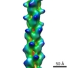 5129MC 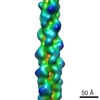 5128C  3ikuC C: citing same article ( M: map data used to model this data |
|---|---|
| Similar structure data |


-Links
 PDBj
PDBj- Assembly
| Deposited unit | 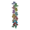
|
|---|---|
| 1 |
|
-Components
| #1: Protein | Mass: 35804.375 Da / Num. of mol.: 12 Source method: isolated from a genetically manipulated source Source: (gene. exp.) |
|---|
-Experimental details
-Experiment
| Experiment | Method: ELECTRON MICROSCOPY |
|---|---|
| EM experiment | Aggregation state: FILAMENT / 3D reconstruction method: helical reconstruction |
- Sample preparation
| Component | Name: ParM Filament in Open State / Type: COMPLEX |
|---|---|
| Specimen | Embedding applied: NO / Shadowing applied: NO / Staining applied: NO / Vitrification applied: YES |
| Vitrification | Cryogen name: ETHANE |
- Electron microscopy imaging
| Experimental equipment |  Model: Tecnai F20 / Image courtesy: FEI Company |
|---|---|
| Microscopy | Model: FEI TECNAI F20 |
| Electron gun | Electron source:  FIELD EMISSION GUN / Accelerating voltage: 200 kV / Illumination mode: FLOOD BEAM FIELD EMISSION GUN / Accelerating voltage: 200 kV / Illumination mode: FLOOD BEAM |
| Electron lens | Mode: BRIGHT FIELD |
| Radiation | Protocol: SINGLE WAVELENGTH / Monochromatic (M) / Laue (L): M |
| Radiation wavelength | Relative weight: 1 |
- Processing
| 3D reconstruction | Method: IHRSR / Resolution: 18 Å / Nominal pixel size: 2.38 Å / Actual pixel size: 2.38 Å / Symmetry type: HELICAL | ||||||||||||
|---|---|---|---|---|---|---|---|---|---|---|---|---|---|
| Refinement step | Cycle: LAST
|
