 ムービー
ムービー コントローラー
 構造ビューア
 EMN検索について


-検索条件
-検索結果
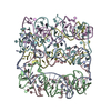
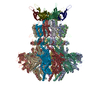
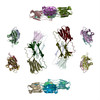
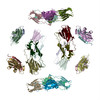
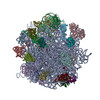
検索 (著者・登録者: e. & v. & orlova)の結果全42件を表示しています

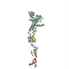
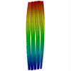
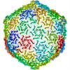
PDB-7ywh: 
Six DNA Helix Bundle nanopore - State 1
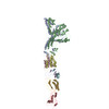
PDB-7ywi:
Six DNA duplex bundle nanopore - State 2

PDB-7ywl:
Six DNA Helix Bundle nanopore - State 3

PDB-7ywn:
Six DNA Helix Bundle nanopore - State 4

PDB-7ywo:
Six DNA Helix Bundle nanopore - State 5
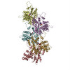
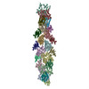
PDB-7z4w:
gp6/gp15/gp16 connector complex of bacteriophage SPP1

PDB-7pv2:
GA1 bacteriophage portal protein

PDB-7pv4:
PhiCPV4 bacteriophage Portal Protein

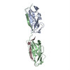
PDB-7apd:
Bovine Papillomavirus E1 DNA helicase-replication fork complex
PDB-6hqe:
Cryo-EM of self-assembly peptide filament LRV_M3delta1

PDB-6mk1:
Cryo-EM of self-assembly peptide filament HEAT_R1
PDB-5a79:
Novel inter-subunit contacts in Barley Stripe Mosaic Virus revealed by cryo-EM
PDB-5a7a:
Novel inter-subunit contacts in Barley Stripe Mosaic Virus revealed by cryo-EM

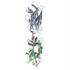
PDB-5a9k:
Structural basis for DNA strand separation by a hexameric replicative helicase
PDB-5a20:
Structure of bacteriophage SPP1 head-to-tail interface filled with DNA and tape measure protein
PDB-5a21:
Structure of bacteriophage SPP1 head-to-tail interface without DNA and tape measure protein
PDB-3j8i:
Near-Atomic Resolution for One State of F-Actin
PDB-3j8j:
Tilted state of actin, T1
PDB-3j8k:
Tilted state of actin, T2
PDB-2m5k:
Atomic-resolution structure of a doublet cross-beta amyloid fibril
PDB-2m5m:
Atomic-resolution structure of a triplet cross-beta amyloid fibril
PDB-3zpk:
Atomic-resolution structure of a quadruplet cross-beta amyloid fibril.
PDB-2ypw:
Atomic model for the N-terminus of TraO fitted in the full-length structure of the bacterial pKM101 type IV secretion system core complex
PDB-3zbi:
Fitting result in the O-layer of the subnanometer structure of the bacterial pKM101 type IV secretion system core complex digested with elastase
PDB-3zbj:
Fitting results in the I-layer of the subnanometer structure of the bacterial pKM101 type IV secretion system core complex digested with elastase
PDB-4an5:
Capsid structure and its Stability at the Late Stages of Bacteriophage SPP1 Assembly
PDB-3j0s:
Remodeling of actin filaments by ADF cofilin proteins

PDB-2xea:
4.6 ANGSTROM CRYO-EM RECONSTRUCTION OF TOBACCO MOSAIC VIRUS FROM IMAGES RECORDED AT 300 KEV ON A 4KX4K CCD CAMERA

PDB-3lue:
Model of alpha-actinin CH1 bound to F-actin

PDB-3iku:
Structural model of ParM filament in closed state from cryo-EM

PDB-3iky:
Structural model of ParM filament in the open state by cryo-EM
PDB-3cre:
Electron Microscopy model of the Saf Pilus- Type A
PDB-3crf:
Electron Microscopy model of the Saf Pilus- Type B

PDB-3byh:
Model of actin-fimbrin ABD2 complex
PDB-2qu4:
Model for Bacterial ParM Filament

PDB-2j9i:
Lengsin is a survivor of an ancient family of class I glutamine synthetases in eukaryotes that has undergone evolutionary re- engineering for a tissue-specific role in the vertebrate eye lens.
PDB-2h50:
Multiple distinct assemblies reveal conformational flexibility in the small heat shock protein Hsp26
PDB-2h53:
Multiple distinct assemblies reveal conformational flexibility in the small heat shock protein Hsp26

PDB-2byu:
Negative stain EM reconstruction of M.tuberculosis Acr1(Hsp 16.3) fitted with wheat sHSP dimer
PDB-2bk1:
The pore structure of pneumolysin, obtained by fitting the alpha carbon trace of perfringolysin O into a cryo-EM map

PDB-2bk2:
The prepore structure of pneumolysin, obtained by fitting the alpha carbon trace of perfringolysin O into a cryo-EM map
PDB-1ml5:
Structure of the E. coli ribosomal termination complex with release factor 2
 wwPDBはEMDBデータモデルのバージョン3へ移行します
