Movie
Movie Controller

[English] 日本語
 Yorodumi
- PDB-2j9i: Lengsin is a survivor of an ancient family of class I glutamine s... -
+ Open data

- Basic information
| Entry | Database: PDB / ID: 2j9i | ||||||
|---|---|---|---|---|---|---|---|
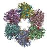
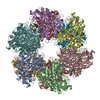
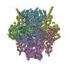
| Title | Lengsin is a survivor of an ancient family of class I glutamine synthetases in eukaryotes that has undergone evolutionary re- engineering for a tissue-specific role in the vertebrate eye lens. | ||||||
 Components Components | GLUTAMATE-AMMONIA LIGASE DOMAIN-CONTAINING PROTEIN 1 | ||||||
 Keywords Keywords |  LIGASE LIGASE | ||||||
| Function / homology |  Function and homology information Function and homology information | ||||||
| Biological species |  MUS MUSCULUS (house mouse) MUS MUSCULUS (house mouse) | ||||||
| Method | ELECTRON MICROSCOPY / single particle reconstruction / cryo EM / Resolution: 17 Å | ||||||
 Authors Authors | Wyatt, K. / White, H.E. / Wang, L. / Bateman, O.A. / Slingsby, C. / Orlova, E.V. / Wistow, G. | ||||||
 Citation Citation | Journal: Structure / Year: 2006 Title: Lengsin is a survivor of an ancient family of class I glutamine synthetases re-engineered by evolution for a role in the vertebrate lens. Authors: Keith Wyatt / Helen E White / Luchun Wang / Orval A Bateman / Christine Slingsby / Elena V Orlova / Graeme Wistow /  Abstract: Lengsin is a major protein of the vertebrate eye lens. It belongs to the hitherto purely prokaryotic GS I branch of the glutamine synthetase (GS) superfamily, but has no enzyme activity. Like the ...Lengsin is a major protein of the vertebrate eye lens. It belongs to the hitherto purely prokaryotic GS I branch of the glutamine synthetase (GS) superfamily, but has no enzyme activity. Like the taxon-specific crystallins, Lengsin is the result of the recruitment of an ancient enzyme to a noncatalytic role in the vertebrate lens. Cryo-EM and modeling studies of Lengsin show a dodecamer structure with important similarities and differences with prokaryotic GS I structures. GS homology regions of Lengsin are well conserved, but the N-terminal domain shows evidence of dynamic evolutionary changes. Compared with birds and fish, most mammals have an additional exon corresponding to part of the N-terminal domain; however, in human, this is a nonfunctional pseudoexon. Genes related to Lengsin are also present in the sea urchin, suggesting that this branch of the GS I family, supplanted by GS II enzymes in vertebrates, has an ancient role in metazoans. | ||||||
| History |
| ||||||
| Remark 700 | SHEET THE SHEET STRUCTURE OF THIS MOLECULE IS BIFURCATED. IN ORDER TO REPRESENT THIS FEATURE IN ... SHEET THE SHEET STRUCTURE OF THIS MOLECULE IS BIFURCATED. IN ORDER TO REPRESENT THIS FEATURE IN THE SHEET RECORDS BELOW, TWO SHEETS ARE DEFINED. |
- Structure visualization
| Movie |
Movie viewer |
|---|---|
| Structure viewer | Molecule: MolmilJmol/JSmol |
- Downloads & links
-Download
| PDBx/mmCIF format | 2j9i.cif.gz | 701 KB | Display | PDBx/mmCIF format |
|---|---|---|---|---|
| PDB format | pdb2j9i.ent.gz | 576.5 KB | Display | PDB format |
| PDBx/mmJSON format | 2j9i.json.gz | Tree view | PDBx/mmJSON format | |
| Others |  Other downloads Other downloads |
-Validation report
| Arichive directory | https://data.pdbj.org/pub/pdb/validation_reports/j9/2j9iftp://data.pdbj.org/pub/pdb/validation_reports/j9/2j9i | HTTPS FTP |
|---|
-Related structure data
| Related structure data |  1290MC M: map data used to model this data C: citing same article ( |
|---|---|
| Similar structure data |


-Links
 PDBj
PDBj
- Assembly
| Deposited unit | 
|
|---|---|
| 1 |
|
-Components
| #1: Protein | Mass: 39588.000 Da / Num. of mol.: 12 Source method: isolated from a genetically manipulated source Source: (gene. exp.) MUS MUSCULUS (house mouse) / Production host:  ESCHERICHIA COLI (E. coli) / References: UniProt: Q8CIX8*PLUS ESCHERICHIA COLI (E. coli) / References: UniProt: Q8CIX8*PLUS |
|---|
-Experimental details
-Experiment
| Experiment | Method: ELECTRON MICROSCOPY |
|---|---|
| EM experiment | Aggregation state: PARTICLE / 3D reconstruction method: single particle reconstruction |
- Sample preparation
| Component | Name: LENGSIN / Type: COMPLEX |
|---|---|
| Buffer solution | Name: 25 MM MALONIC ACID / pH: 6 / Details: 25 MM MALONIC ACID |
| Specimen | Conc.: 0.3 mg/ml / Embedding applied: NO / Shadowing applied: NO / Staining applied: NO / Vitrification applied: YES |
| Specimen support | Details: HOLEY CARBON |
| Vitrification | Cryogen name: ETHANE |
- Electron microscopy imaging
| Experimental equipment |  Model: Tecnai F20 / Image courtesy: FEI Company |
|---|---|
| Microscopy | Model: FEI TECNAI F20 |
| Electron gun | Electron source: FIELD EMISSION GUN / Accelerating voltage: 200 kV / Illumination mode: FLOOD BEAM |
| Electron lens | Mode: BRIGHT FIELDBright-field microscopy / Nominal magnification: 50000 X / Nominal defocus max: 320 nm / Nominal defocus min: 170 nm |
| Image recording | Film or detector model: KODAK SO-163 FILM |
| Radiation wavelength | Relative weight: 1 |
- Processing
| EM software |
| ||||||||||||
|---|---|---|---|---|---|---|---|---|---|---|---|---|---|
| CTF correction | Details: PHASE FLIPPING | ||||||||||||
| Symmetry | Point symmetry: I (icosahedral) | ||||||||||||
| 3D reconstruction | Method: EXACT-FILTER BACK PROJECTION / Resolution: 17 Å / Nominal pixel size: 1.4 Å / Actual pixel size: 1.45 Å Details: THE HOMOLOGY MODEL WAS MINIMIZED IN AMBER 8 PRIOR TO MANUAL FITTING Symmetry type: POINT | ||||||||||||
| Atomic model building | Protocol: OTHER / Details: METHOD--URO | ||||||||||||
| Atomic model building | PDB-ID: 1F52 | ||||||||||||
| Refinement | Highest resolution: 17 Å | ||||||||||||
| Refinement step | Cycle: LAST / Highest resolution: 17 Å
|