Movie
Movie Controller


+ Open data

- Basic information
| Entry | Database: PDB / ID: 4a7h | ||||||
|---|---|---|---|---|---|---|---|
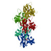
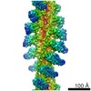
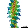
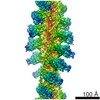
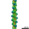
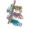
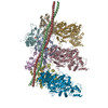
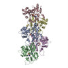
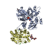
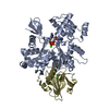
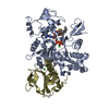
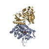
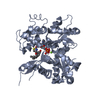
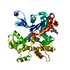
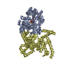
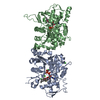
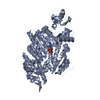
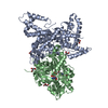
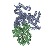
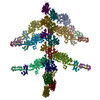
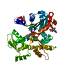
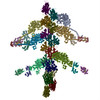
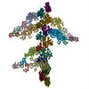
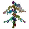
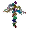
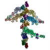
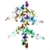
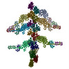
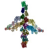
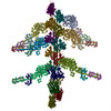
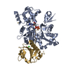
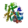
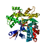
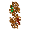
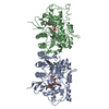
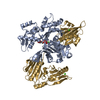
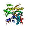
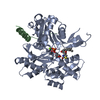
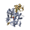
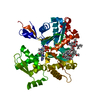
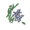
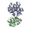
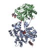
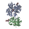
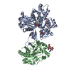
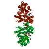
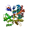
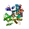
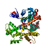
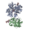
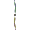
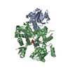
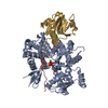
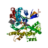
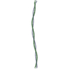
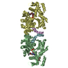
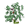
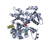
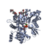
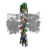
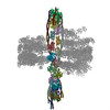
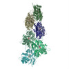
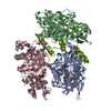
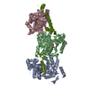
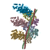
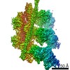
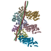
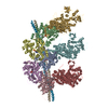
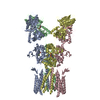
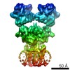
| Title | Structure of the Actin-Tropomyosin-Myosin Complex (rigor ATM 2) | ||||||
 Components Components |
| ||||||
 Keywords Keywords | STRUCTURAL PROTEIN/HYDROLASE / STRUCTURAL PROTEIN-HYDROLASE COMPLEX / STRUCTURAL PROTEIN / CYTOSKELETON / CONTRACTILE FILAMENT / MOTOR ACTIVITY / MYOSIN BINDING / ACTIN BINDING / ATP CATABOLIC PROCESS / RIGOR STATE | ||||||
| Function / homology |  Function and homology information Function and homology informationmacropinocytic cup membrane / pseudopodium membrane / actin wave / macropinocytic cup cytoskeleton / chemotaxis to cAMP / macropinocytic cup / actin filament-based movement / leading edge membrane / early phagosome / myosin complex ...macropinocytic cup membrane / pseudopodium membrane / actin wave / macropinocytic cup cytoskeleton / chemotaxis to cAMP / macropinocytic cup / actin filament-based movement / leading edge membrane / early phagosome / myosin complex / cytoskeletal motor activator activity / microfilament motor activity / phagocytosis, engulfment / tropomyosin binding / mesenchyme migration / troponin I binding / myosin heavy chain binding / cell leading edge / pseudopodium / filamentous actin / actin filament bundle / phosphatidylinositol-3,4,5-trisphosphate binding / skeletal muscle thin filament assembly / striated muscle thin filament / actin filament bundle assembly / phagocytic cup / skeletal muscle myofibril / actin monomer binding / skeletal muscle fiber development / phagocytosis / muscle contraction / stress fiber / titin binding / actin filament polymerization / filopodium / actin filament organization / actin filament / Hydrolases; Acting on acid anhydrides; Acting on acid anhydrides to facilitate cellular and subcellular movement / actin filament binding / endocytosis / calcium-dependent protein binding / actin cytoskeleton / lamellipodium / actin binding / cell body / calmodulin binding / hydrolase activity / protein domain specific binding / protein heterodimerization activity / calcium ion binding / positive regulation of gene expression / magnesium ion binding / protein homodimerization activity / ATP binding / identical protein binding / plasma membrane / cytoplasm / cytosol Similarity search - Function | ||||||
| Biological species |   | ||||||
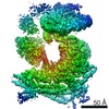
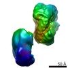
| Method | ELECTRON MICROSCOPY / helical reconstruction / cryo EM / Resolution: 7.8 Å | ||||||
 Authors Authors | Behrmann, E. / Mueller, M. / Penczek, P.A. / Mannherz, H.G. / Manstein, D.J. / Raunser, S. | ||||||
 Citation Citation | Journal: Cell / Year: 2012 Title: Structure of the rigor actin-tropomyosin-myosin complex. Authors: Elmar Behrmann / Mirco Müller / Pawel A Penczek / Hans Georg Mannherz / Dietmar J Manstein / Stefan Raunser /  Abstract: Regulation of myosin and filamentous actin interaction by tropomyosin is a central feature of contractile events in muscle and nonmuscle cells. However, little is known about molecular interactions ...Regulation of myosin and filamentous actin interaction by tropomyosin is a central feature of contractile events in muscle and nonmuscle cells. However, little is known about molecular interactions within the complex and the trajectory of tropomyosin movement between its "open" and "closed" positions on the actin filament. Here, we report the 8 Å resolution structure of the rigor (nucleotide-free) actin-tropomyosin-myosin complex determined by cryo-electron microscopy. The pseudoatomic model of the complex, obtained from fitting crystal structures into the map, defines the large interface involving two adjacent actin monomers and one tropomyosin pseudorepeat per myosin contact. Severe forms of hereditary myopathies are linked to mutations that critically perturb this interface. Myosin binding results in a 23 Å shift of tropomyosin along actin. Complex domain motions occur in myosin, but not in actin. Based on our results, we propose a structural model for the tropomyosin-dependent modulation of myosin binding to actin. | ||||||
| History |
|
- Structure visualization
| Movie |
Movie viewer |
|---|---|
| Structure viewer | Molecule: MolmilJmol/JSmol |
- Downloads & links
-Download
| PDBx/mmCIF format | 4a7h.cif.gz | 761.3 KB | Display | PDBx/mmCIF format |
|---|---|---|---|---|
| PDB format | pdb4a7h.ent.gz | 631.6 KB | Display | PDB format |
| PDBx/mmJSON format | 4a7h.json.gz | Tree view | PDBx/mmJSON format | |
| Others |  Other downloads Other downloads |
-Validation report
| Summary document | 4a7h_validation.pdf.gz | 1.1 MB | Display | wwPDB validaton report |
|---|---|---|---|---|
| Full document | 4a7h_full_validation.pdf.gz | 1.2 MB | Display | |
| Data in XML | 4a7h_validation.xml.gz | 118 KB | Display | |
| Data in CIF | 4a7h_validation.cif.gz | 168.4 KB | Display | |
| Arichive directory | https://data.pdbj.org/pub/pdb/validation_reports/a7/4a7hftp://data.pdbj.org/pub/pdb/validation_reports/a7/4a7h | HTTPS FTP |
-Related structure data


-Links
 PDBj
PDBj


- Assembly
| Deposited unit | 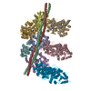
|
|---|---|
| 1 |
|
-Components
| #1: Protein | Mass: 41875.633 Da / Num. of mol.: 5 / Source method: isolated from a natural source / Source: (natural) #2: Protein | Mass: 15807.628 Da / Num. of mol.: 2 / Fragment: RESIDUES 98-233 Source method: isolated from a genetically manipulated source Source: (gene. exp.)  #3: Protein | Mass: 79207.305 Da / Num. of mol.: 3 / Fragment: RESIDUES 1-697 / Mutation: YES Source method: isolated from a genetically manipulated source Source: (gene. exp.) #4: Chemical | ChemComp-ADP /   Mass: 427.201 Da / Num. of mol.: 5 / Source method: obtained synthetically / Formula: C10H15N5O10P2 / Comment: ADP, energy-carrying molecule*YM Mass: 427.201 Da / Num. of mol.: 5 / Source method: obtained synthetically / Formula: C10H15N5O10P2 / Comment: ADP, energy-carrying molecule*YM#5: Chemical | ChemComp-CA /   Mass: 40.078 Da / Num. of mol.: 5 / Source method: obtained synthetically / Formula: Ca Mass: 40.078 Da / Num. of mol.: 5 / Source method: obtained synthetically / Formula: CaCompound details | ENGINEERED RESIDUE IN CHAIN C, SER 334 TO GLU ENGINEERED RESIDUE IN CHAIN I, SER 334 TO GLU ...ENGINEERED | Sequence details | SEQUENCE IS NOT BASED ON THE EXPERIMENTAL PROTEIN AS NO FULL-LENGTH TROPOMYOSIN STRUCTURES WERE ...SEQUENCE IS NOT BASED ON THE EXPERIMENT | |
|---|
-Experimental details
-Experiment
| Experiment | Method: ELECTRON MICROSCOPY |
|---|---|
| EM experiment | Aggregation state: FILAMENT / 3D reconstruction method: helical reconstruction |
- Sample preparation
| Component | Name: F-ACTIN-MYO1E-TROPOMYOSIN COMPLEX (CONFORMATION 2) / Type: COMPLEX / Details: CMOS IMAGE FRAMES SELECTED BY POWER SPECTRUM |
|---|---|
| Buffer solution | Name: 5MM TRIS, 100MM KCL, 2MM MGCL2, 50MM GLUTAMINE, 50MM ARGININ pH: 7.2 Details: 5MM TRIS, 100MM KCL, 2MM MGCL2, 50MM GLUTAMINE, 50MM ARGININ |
| Specimen | Conc.: 0.01 mg/ml / Embedding applied: NO / Shadowing applied: NO / Staining applied: NO / Vitrification applied: YES |
| Specimen support | Details: HOLEY CARBON |
| Vitrification | Instrument: GATAN CRYOPLUNGE 3 / Cryogen name: ETHANE Details: VITRIFICATION 1 -- CRYOGEN- ETHANE, HUMIDITY- 90, TEMPERATURE- 101, INSTRUMENT- GATAN CRYOPLUNGE 3, METHOD- MANUAL BLOTTING FOR APPROXIMATELY 15 SECONDS, |
- Electron microscopy imaging
| Microscopy | Model: JEOL 3200FSC Details: BEST 836 MICROGRAPHS WERE SELECTED FROM OVER 3000 AQUIRED IMAGES |
|---|---|
| Electron gun | Electron source:  FIELD EMISSION GUN / Accelerating voltage: 200 kV / Illumination mode: FLOOD BEAM FIELD EMISSION GUN / Accelerating voltage: 200 kV / Illumination mode: FLOOD BEAM |
| Electron lens | Mode: BRIGHT FIELD / Nominal magnification: 80000 X / Calibrated magnification: 169644 X / Nominal defocus max: 1500 nm / Nominal defocus min: 750 nm / Cs: 4.1 mm |
| Specimen holder | Temperature: 77 K |
| Image recording | Electron dose: 1.7 e/Å2 / Film or detector model: TVIPS TEMCAM-F816 (8k x 8k) |
| Image scans | Num. digital images: 836 |
| Radiation wavelength | Relative weight: 1 |
- Processing
| EM software |
| ||||||||||||
|---|---|---|---|---|---|---|---|---|---|---|---|---|---|
| CTF correction | Details: EACH PARTICLE | ||||||||||||
| 3D reconstruction | Method: IHRSR / Resolution: 7.8 Å / Num. of particles: 9650 / Nominal pixel size: 1.84 Å / Actual pixel size: 1.84 Å Details: SUBMISSION BASED ON EXPERIMENTAL DATA FROM EMDB EMD-1988 (DEPOSITION ID: 10379). Symmetry type: HELICAL | ||||||||||||
| Atomic model building | Protocol: FLEXIBLE FIT / Space: REAL Details: METHOD--GEOMETRY-BASED CONFORMATIONAL SAMPLING USING DEFORMABLE ELASTIC NETWORK (DEN) APPROACH REFINEMENT PROTOCOL--EM | ||||||||||||
| Atomic model building |
| ||||||||||||
| Refinement | Highest resolution: 7.8 Å | ||||||||||||
| Refinement step | Cycle: LAST / Highest resolution: 7.8 Å
|