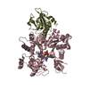
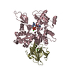
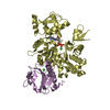
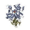
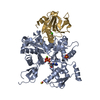
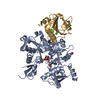
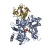
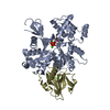
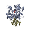
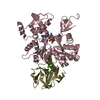
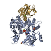
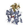
SEQUENCE FUSION OF GELSOLIN DOMAIN 1 FROM HUMAN WITH C-TERMINAL DOMAIN OF THYMOSIN B4 FROM MOUSE ...SEQUENCE FUSION OF GELSOLIN DOMAIN 1 FROM HUMAN WITH C-TERMINAL DOMAIN OF THYMOSIN B4 FROM MOUSE (SEQUENCE:ETQEKNPLPSKETIEQEKQ)
Resolution: 2→20 Å / Cor.coef. Fo:Fc: 0.963 / Cor.coef. Fo:Fc free: 0.937 / SU B: 2.957 / SU ML: 0.085 / Cross valid method: THROUGHOUT / ESU R Free: 0.134 / Details: HYDROGENS HAVE BEEN ADDED IN THE RIDING POSITIONS
Rfactor
Num. reflection
% reflection
Selection details
Rfree
0.19531
2079
5 %
RANDOM
Rwork
0.14445
-
-
-
obs
0.14709
39223
98.15 %
-
Solvent computation
Ion probe radii: 0.8 Å / Shrinkage radii: 0.8 Å / VDW probe radii: 1.4 Å / Solvent model: BABINET MODEL WITH MASK
Displacement parameters
Biso mean: 14.977 Å2
Baniso -1
Baniso -2
Baniso -3
1-
1.14 Å2
0 Å2
0.35 Å2
2-
-
-0.42 Å2
0 Å2
3-
-
-
-0.66 Å2
Refinement step
Cycle: LAST / Resolution: 2→20 Å
Protein
Nucleic acid
Ligand
Solvent
Total
Num. atoms
3959
0
34
427
4420
Refine LS restraints
Refine-ID
Type
Dev ideal
Dev ideal target
Number
X-RAY DIFFRACTION
r_bond_refined_d
0.014
0.021
4082
X-RAY DIFFRACTION
r_bond_other_d
0.002
0.02
3627
X-RAY DIFFRACTION
r_angle_refined_deg
1.434
1.963
5535
X-RAY DIFFRACTION
r_angle_other_deg
0.868
3
8473
X-RAY DIFFRACTION
r_dihedral_angle_1_deg
5.574
5
499
X-RAY DIFFRACTION
r_chiral_restr
0.094
0.2
603
X-RAY DIFFRACTION
r_gen_planes_refined
0.005
0.02
4512
X-RAY DIFFRACTION
r_gen_planes_other
0.004
0.02
809
X-RAY DIFFRACTION
r_nbd_refined
0.203
0.2
784
X-RAY DIFFRACTION
r_nbd_other
0.245
0.2
4127
X-RAY DIFFRACTION
r_nbtor_other
0.086
0.2
2242
X-RAY DIFFRACTION
r_xyhbond_nbd_refined
0.162
0.2
304
X-RAY DIFFRACTION
r_metal_ion_refined
0.078
0.2
15
X-RAY DIFFRACTION
r_symmetry_vdw_refined
0.068
0.2
3
X-RAY DIFFRACTION
r_symmetry_vdw_other
0.252
0.2
42
X-RAY DIFFRACTION
r_symmetry_hbond_refined
0.106
0.2
19
X-RAY DIFFRACTION
r_mcbond_it
0.969
1.5
2494
X-RAY DIFFRACTION
r_mcangle_it
1.629
2
4025
X-RAY DIFFRACTION
r_scbond_it
2.95
3
1588
X-RAY DIFFRACTION
r_scangle_it
4.545
4.5
1510
LS refinement shell
Resolution: 2→2.051 Å / Total num. of bins used: 20 /
Rfactor
Num. reflection
Rfree
0.196
140
Rwork
0.127
2890
Refinement TLS params.
Method: refined / Refine-ID: X-RAY DIFFRACTION
ID
L11 (°2)
L12 (°2)
L13 (°2)
L22 (°2)
L23 (°2)
L33 (°2)
S11 (Å °)
S12 (Å °)
S13 (Å °)
S21 (Å °)
S22 (Å °)
S23 (Å °)
S31 (Å °)
S32 (Å °)
S33 (Å °)
T11 (Å2)
T12 (Å2)
T13 (Å2)
T22 (Å2)
T23 (Å2)
T33 (Å2)
Origin x (Å)
Origin y (Å)
Origin z (Å)
1
0.5605
0.1648
-0.2891
1.118
0.1821
1.1494
-0.019
-0.0111
-0.0442
-0.0277
0.0356
-0.1174
0.0297
0.0954
-0.0166
0.0634
-0.0043
0.0115
0.1292
-0.0121
0.1194
13.27
-8.442
23.034
2
3.4305
1.4418
-0.6866
8.0116
-1.3148
2.9583
0.1536
0.1717
0.0814
-0.1824
-0.0247
-0.3562
0.0883
0.2529
-0.1289
0.0516
-0.0136
0.06
0.1358
-0.0263
0.0745
18.528
1.199
3.708
3
0.8275
-0.0942
0.0683
0.7237
-0.1121
1.2282
-0.0133
0.0317
0.0173
-0.0227
0.0288
0.0389
0.0466
-0.0964
-0.0155
0.0833
-0.0118
0.0025
0.1425
0.0092
0.1016
-10.515
-0.451
22.771
4
0.9088
-0.1035
-0.3906
1.1919
-0.3899
1.0628
0.0332
0.0584
0.0367
-0.1388
-0.0017
-0.0331
-0.01
-0.0762
-0.0315
0.1104
-0.0016
0.0049
0.1341
0.0351
0.0662
-4.781
13.529
3.125
5
0.6797
0.4544
-0.2006
1.5906
-0.0675
0.9111
0.0099
-0.0591
-0.0098
0.1737
0.0096
0.0193
0.013
0.0458
-0.0195
0.1344
0.0056
0.0027
0.1038
0.0003
0.0792
-2.812
-1.786
47.438
6
1.0654
0.3132
-1
0.9998
-4.8265
32.7026
0.1943
0.1473
0.3959
0.0083
-0.016
-0.2443
-0.0383
0.3091
-0.1783
0.094
0.0203
0.0962
0.0756
0.052
0.157
15.596
12.659
3.664
7
0.0662
-0.0219
-0.1275
0.2688
-0.0315
0.3065
0.0024
0.0201
0.0092
0.0119
0.0066
-0.01
0.0121
-0.0104
-0.009
0.1027
-0.0148
-0.0028
0.128
0.0088
0.1012
-1.004
1.347
25.051
Refinement TLS group
ID
Refine-ID
Refine TLS-ID
Auth asym-ID
Label asym-ID
Auth seq-ID
Label seq-ID
1
X-RAY DIFFRACTION
1
A
B
6 - 32
1 - 27
2
X-RAY DIFFRACTION
1
A
B
71 - 136
66 - 131
3
X-RAY DIFFRACTION
1
A
B
337 - 375
332 - 370
4
X-RAY DIFFRACTION
1
A
C
700
1
5
X-RAY DIFFRACTION
2
A
B
33 - 70
28 - 65
6
X-RAY DIFFRACTION
3
A
B
137 - 180
132 - 175
7
X-RAY DIFFRACTION
3
A
B
274 - 336
269 - 331
8
X-RAY DIFFRACTION
4
A
B
181 - 273
176 - 268
9
X-RAY DIFFRACTION
4
A
F
900
1
10
X-RAY DIFFRACTION
5
G
A
28 - 152
4 - 128
11
X-RAY DIFFRACTION
5
G
D - E
701 - 702
1
12
X-RAY DIFFRACTION
6
G
A
153 - 171
129 - 147
13
X-RAY DIFFRACTION
7
A
G
901 - 1175
1 - 275
+
About Yorodumi
-
News
-
Feb 9, 2022. New format data for meta-information of EMDB entries
New format data for meta-information of EMDB entries
Version 3 of the EMDB header file is now the official format.
The previous official version 1.9 will be removed from the archive.
In the structure databanks used in Yorodumi, some data are registered as the other names, "COVID-19 virus" and "2019-nCoV". Here are the details of the virus and the list of structure data.
Jan 31, 2019. EMDB accession codes are about to change! (news from PDBe EMDB page)
EMDB accession codes are about to change! (news from PDBe EMDB page)
The allocation of 4 digits for EMDB accession codes will soon come to an end. Whilst these codes will remain in use, new EMDB accession codes will include an additional digit and will expand incrementally as the available range of codes is exhausted. The current 4-digit format prefixed with “EMD-” (i.e. EMD-XXXX) will advance to a 5-digit format (i.e. EMD-XXXXX), and so on. It is currently estimated that the 4-digit codes will be depleted around Spring 2019, at which point the 5-digit format will come into force.
The EM Navigator/Yorodumi systems omit the EMD- prefix.
Related info.:Q: What is EMD? / ID/Accession-code notation in Yorodumi/EM Navigator
Yorodumi is a browser for structure data from EMDB, PDB, SASBDB, etc.
This page is also the successor to EM Navigator detail page, and also detail information page/front-end page for Omokage search.
The word "yorodu" (or yorozu) is an old Japanese word meaning "ten thousand". "mi" (miru) is to see.
Related info.:EMDB / PDB / SASBDB / Comparison of 3 databanks / Yorodumi Search / Aug 31, 2016. New EM Navigator & Yorodumi / Yorodumi Papers / Jmol/JSmol / Function and homology information / Changes in new EM Navigator and Yorodumi
 Movie
Movie Controller

 Yorodumi
 Open data
 Basic information
 Components
 Keywords
 STRUCTURAL PROTEIN
 Function and homology information

 Authors
 Citation
 Structure visualization
 Downloads & links
 Other downloads
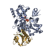


 PDBj
PDBj


 Assembly
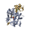


 Mass: 40.078 Da / Num. of mol.: 3 / Source method: obtained synthetically / Formula: Ca
 Mass: 507.181 Da / Num. of mol.: 1 / Source method: obtained synthetically / Formula: C10H16N5O13P3 / Comment: ATP, energy-carrying molecule*YM
 Mass: 18.015 Da / Num. of mol.: 427 / Source method: isolated from a natural source / Formula: H2O
 Sample preparation
 / Beamline: ID29 / Wavelength: 1.0052 Å
 Processing