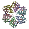
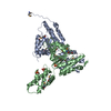
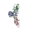
Journal: Nature / Year: 2011 Title: Structure and function of the AAA+ protein CbbX, a red-type Rubisco activase. Authors: Oliver Mueller-Cajar / Mathias Stotz / Petra Wendler / F Ulrich Hartl / Andreas Bracher / Manajit Hayer-Hartl / Abstract: Ribulose 1,5-bisphosphate carboxylase/oxygenase (Rubisco) catalyses the fixation of atmospheric CO(2) in photosynthesis, but tends to form inactive complexes with its substrate ribulose 1,5- ...Ribulose 1,5-bisphosphate carboxylase/oxygenase (Rubisco) catalyses the fixation of atmospheric CO(2) in photosynthesis, but tends to form inactive complexes with its substrate ribulose 1,5-bisphosphate (RuBP). In plants, Rubisco is reactivated by the AAA(+) (ATPases associated with various cellular activities) protein Rubisco activase (Rca), but no such protein is known for the Rubisco of red algae. Here we identify the protein CbbX as an activase of red-type Rubisco. The 3.0-Å crystal structure of unassembled CbbX from Rhodobacter sphaeroides revealed an AAA(+) protein architecture. Electron microscopy and biochemical analysis showed that ATP and RuBP must bind to convert CbbX into functionally active, hexameric rings. The CbbX ATPase is strongly stimulated by RuBP and Rubisco. Mutational analysis suggests that CbbX functions by transiently pulling the carboxy-terminal peptide of the Rubisco large subunit into the hexamer pore, resulting in the release of the inhibitory RuBP. Understanding Rubisco activation may facilitate efforts to improve CO(2) uptake and biomass production by photosynthetic organisms.
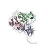
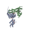
THE ANALYSIS OF THE CBBX PROTEIN IN SOLUTION AND EM STUDIES SUGGEST THAT THE BIOLOGICALLY ACTIVE OLIGOMER IS A HEXAMER, BUT IT CANNOT BE GENERATED BY THE APPLICATION OF SYMMETRY OPERATORS TO THE CHAINS IN THE COORDINATE FILE.
-
Components
#1: Protein
ProteinCbbX
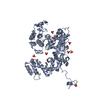
Mass: 34717.289 Da / Num. of mol.: 2 / Mutation: M282I Source method: isolated from a genetically manipulated source Source: (gene. exp.) Rhodobacter sphaeroides (bacteria) / Strain: KD131 / KCTC 12085 / Gene: cbbX, RSKD131_2679 / Plasmid: pHUE / Production host: Escherichia coli (E. coli) / Strain (production host): BL21(DE3) / References: UniProt: P95648
In the structure databanks used in Yorodumi, some data are registered as the other names, "COVID-19 virus" and "2019-nCoV". Here are the details of the virus and the list of structure data.
Jan 31, 2019. EMDB accession codes are about to change! (news from PDBe EMDB page)
EMDB accession codes are about to change! (news from PDBe EMDB page)
The allocation of 4 digits for EMDB accession codes will soon come to an end. Whilst these codes will remain in use, new EMDB accession codes will include an additional digit and will expand incrementally as the available range of codes is exhausted. The current 4-digit format prefixed with “EMD-” (i.e. EMD-XXXX) will advance to a 5-digit format (i.e. EMD-XXXXX), and so on. It is currently estimated that the 4-digit codes will be depleted around Spring 2019, at which point the 5-digit format will come into force.
The EM Navigator/Yorodumi systems omit the EMD- prefix.
Related info.:Q: What is EMD? / ID/Accession-code notation in Yorodumi/EM Navigator
Yorodumi is a browser for structure data from EMDB, PDB, SASBDB, etc.
This page is also the successor to EM Navigator detail page, and also detail information page/front-end page for Omokage search.
The word "yorodu" (or yorozu) is an old Japanese word meaning "ten thousand". "mi" (miru) is to see.
Related info.:EMDB / PDB / SASBDB / Comparison of 3 databanks / Yorodumi Search / Aug 31, 2016. New EM Navigator & Yorodumi / Yorodumi Papers / Jmol/JSmol / Function and homology information / Changes in new EM Navigator and Yorodumi
 Movie
Movie Controller


 Open data
 Basic information
 Components
 Keywords
 Function and homology information
 Rhodobacter sphaeroides (bacteria)
 X-RAY DIFFRACTION /
  Authors
 Citation
 Structure visualization
 Downloads & links
 Other downloads
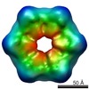

 PDBj
PDBj
 Assembly

 Mass: 96.063 Da / Num. of mol.: 6 / Source method: obtained synthetically / Formula: SO4
 Mass: 18.015 Da / Num. of mol.: 23 / Source method: isolated from a natural source / Formula: H2O
 Sample preparation
 / Beamline: ID29 / Wavelength: 1 Å
 Processing