multi-ciliated epithelial cell differentiation / Rb-E2F complex / negative regulation of fat cell proliferation / regulation of DNA biosynthetic process / centriole assembly / motile cilium assembly / Inhibition of replication initiation of damaged DNA by RB1/E2F1 / Transcription of E2F targets under negative control by DREAM complex / Transcription of E2F targets under negative control by p107 (RBL1) and p130 (RBL2) in complex with HDAC1 / cell volume homeostasis ...multi-ciliated epithelial cell differentiation / Rb-E2F complex / negative regulation of fat cell proliferation / regulation of DNA biosynthetic process / centriole assembly / motile cilium assembly / Inhibition of replication initiation of damaged DNA by RB1/E2F1 / Transcription of E2F targets under negative control by DREAM complex / Transcription of E2F targets under negative control by p107 (RBL1) and p130 (RBL2) in complex with HDAC1 / cell volume homeostasis / blood circulation / Activation of NOXA and translocation to mitochondria / anoikis / Activation of PUMA and translocation to mitochondria / DNA-binding transcription activator activity / G1/S-Specific Transcription / epithelial cell development / Transcriptional Regulation by E2F6 / Defective binding of RB1 mutants to E2F1,(E2F2, E2F3) / G0 and Early G1 / positive regulation of G1/S transition of mitotic cell cycle / epidermis development / cis-regulatory region sequence-specific DNA binding / Cyclin E associated events during G1/S transition / Cyclin A:Cdk2-associated events at S phase entry / TP53 Regulates Transcription of Genes Involved in G2 Cell Cycle Arrest / promoter-specific chromatin binding / animal organ morphogenesis / SMAD2/SMAD3:SMAD4 heterotrimer regulates transcription / Oncogene Induced Senescence / Pre-NOTCH Transcription and Translation / Transcriptional regulation of granulopoiesis / RNA polymerase II transcription regulator complex / Cyclin D associated events in G1 / positive regulation of DNA-binding transcription factor activity / sequence-specific double-stranded DNA binding / regulation of cell population proliferation / DNA-binding transcription activator activity, RNA polymerase II-specific / Oxidative Stress Induced Senescence / DNA-binding transcription factor binding / transcription by RNA polymerase II / protein dimerization activity / DNA-binding transcription factor activity, RNA polymerase II-specific / cell cycle / RNA polymerase II cis-regulatory region sequence-specific DNA binding / DNA-binding transcription factor activity / protein domain specific binding / chromatin / regulation of DNA-templated transcription / regulation of transcription by RNA polymerase II / positive regulation of transcription by RNA polymerase II / DNA binding / nucleoplasm / nucleus / cytosol / cytoplasm Similarity search - Function
In the structure databanks used in Yorodumi, some data are registered as the other names, "COVID-19 virus" and "2019-nCoV". Here are the details of the virus and the list of structure data.
Jan 31, 2019. EMDB accession codes are about to change! (news from PDBe EMDB page)
EMDB accession codes are about to change! (news from PDBe EMDB page)
The allocation of 4 digits for EMDB accession codes will soon come to an end. Whilst these codes will remain in use, new EMDB accession codes will include an additional digit and will expand incrementally as the available range of codes is exhausted. The current 4-digit format prefixed with “EMD-” (i.e. EMD-XXXX) will advance to a 5-digit format (i.e. EMD-XXXXX), and so on. It is currently estimated that the 4-digit codes will be depleted around Spring 2019, at which point the 5-digit format will come into force.
The EM Navigator/Yorodumi systems omit the EMD- prefix.
Related info.:Q: What is EMD? / ID/Accession-code notation in Yorodumi/EM Navigator
Yorodumi is a browser for structure data from EMDB, PDB, SASBDB, etc.
This page is also the successor to EM Navigator detail page, and also detail information page/front-end page for Omokage search.
The word "yorodu" (or yorozu) is an old Japanese word meaning "ten thousand". "mi" (miru) is to see.
Related info.:EMDB / PDB / SASBDB / Comparison of 3 databanks / Yorodumi Search / Aug 31, 2016. New EM Navigator & Yorodumi / Yorodumi Papers / Jmol/JSmol / Function and homology information / Changes in new EM Navigator and Yorodumi
 Movie
Movie Controller

 Yorodumi
 Open data
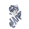
 Basic information
 Components
 Keywords
 TRANSCRIPTION /
  Function and homology information
 Authors
 United States, 1items
  Citation
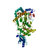
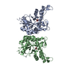
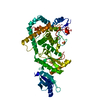
 Structure visualization
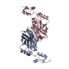
 Downloads & links
 Other downloads

 PDBj
PDBj


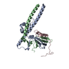
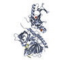
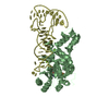
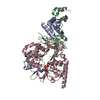
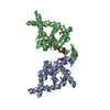
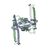
 Assembly


 Mass: 92.094 Da / Num. of mol.: 2 / Source method: obtained synthetically / Formula: C3H8O3
 Mass: 18.015 Da / Num. of mol.: 42 / Source method: isolated from a natural source / Formula: H2O
 Sample preparation
 Processing