SHEET THE SHEET STRUCTURE OF THIS MOLECULE IS BIFURCATED. IN ORDER TO REPRESENT THIS FEATURE IN ... SHEET THE SHEET STRUCTURE OF THIS MOLECULE IS BIFURCATED. IN ORDER TO REPRESENT THIS FEATURE IN THE SHEET RECORDS BELOW, TWO SHEETS ARE DEFINED.
Mass: 18.015 Da / Num. of mol.: 155 / Source method: isolated from a natural source / Formula: H2O
Compound details
ENGINEERED RESIDUE IN CHAIN B, GLY 37 TO ASN ENGINEERED RESIDUE IN CHAIN C, GLY 37 TO ASN ...ENGINEERED RESIDUE IN CHAIN B, GLY 37 TO ASN ENGINEERED RESIDUE IN CHAIN C, GLY 37 TO ASN ENGINEERED RESIDUE IN CHAIN B, THR 123 TO LEU ENGINEERED RESIDUE IN CHAIN C, THR 123 TO LEU
Sequence details
SEQUENCE OF MATURE PROTEIN AFTER SIGNAL PEPTIDE CLIVAGE
-
Experimental details
-
Experiment
Experiment
Method: X-RAY DIFFRACTION / Number of used crystals: 1
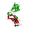
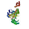
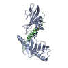
Resolution: 2.5→76.3 Å / Data cutoff high absF: 10000 / Isotropic thermal model: RESTRAINED / Cross valid method: THROUGHOUT / σ(F): 0 / Stereochemistry target values: MAXIMUM LIKELIHOOD Details: THERE ARE 2 MOLECULES OF PAPA IN THE TERNARY COMPLEX (DA2). PAPA MOL B IS DONOR STRAND COMPLEMENTED BY PAPD, PAPA MOL C IS DONOR STRAND EXCHANGED BY PAPA MOL B
Rfactor
Num. reflection
% reflection
Selection details
Rfree
0.2616
1300
5.1 %
RANDOM
Rwork
0.2219
-
-
-
obs
0.2219
25532
99.5 %
-
Solvent computation
Bsol: 46.4546 Å2 / ksol: 0.362486 e/Å3
Displacement parameters
Baniso -1
Baniso -2
Baniso -3
1-
-6.381 Å2
0 Å2
7.304 Å2
2-
-
-0.544 Å2
0 Å2
3-
-
-
6.924 Å2
Refinement step
Cycle: LAST / Resolution: 2.5→76.3 Å
Protein
Nucleic acid
Ligand
Solvent
Total
Num. atoms
3740
0
0
155
3895
Refine LS restraints
Refine-ID
Type
Dev ideal
X-RAY DIFFRACTION
c_bond_d
0.006205
X-RAY DIFFRACTION
c_bond_d_na
X-RAY DIFFRACTION
c_bond_d_prot
X-RAY DIFFRACTION
c_angle_d
X-RAY DIFFRACTION
c_angle_d_na
X-RAY DIFFRACTION
c_angle_d_prot
X-RAY DIFFRACTION
c_angle_deg
1.30842
X-RAY DIFFRACTION
c_angle_deg_na
X-RAY DIFFRACTION
c_angle_deg_prot
X-RAY DIFFRACTION
c_dihedral_angle_d
X-RAY DIFFRACTION
c_dihedral_angle_d_na
X-RAY DIFFRACTION
c_dihedral_angle_d_prot
X-RAY DIFFRACTION
c_improper_angle_d
X-RAY DIFFRACTION
c_improper_angle_d_na
X-RAY DIFFRACTION
c_improper_angle_d_prot
X-RAY DIFFRACTION
c_mcbond_it
X-RAY DIFFRACTION
c_mcangle_it
X-RAY DIFFRACTION
c_scbond_it
X-RAY DIFFRACTION
c_scangle_it
LS refinement shell
Resolution: 2.5→2.53 Å / Total num. of bins used: 26
Rfactor
Num. reflection
% reflection
Rfree
0.345
-
-
Rwork
0.329
863
-
obs
-
-
99.5 %
Xplor file
Refine-ID
Serial no
Param file
Topol file
X-RAY DIFFRACTION
1
PROTEIN_REP.PARAM
PROTEIN.TOP
X-RAY DIFFRACTION
2
WATER_REP.PARAM
WATER.TOP
X-RAY DIFFRACTION
3
ION.PARAM
ION.TOP
+
About Yorodumi
-
News
-
Feb 9, 2022. New format data for meta-information of EMDB entries
New format data for meta-information of EMDB entries
Version 3 of the EMDB header file is now the official format.
The previous official version 1.9 will be removed from the archive.
In the structure databanks used in Yorodumi, some data are registered as the other names, "COVID-19 virus" and "2019-nCoV". Here are the details of the virus and the list of structure data.
Jan 31, 2019. EMDB accession codes are about to change! (news from PDBe EMDB page)
EMDB accession codes are about to change! (news from PDBe EMDB page)
The allocation of 4 digits for EMDB accession codes will soon come to an end. Whilst these codes will remain in use, new EMDB accession codes will include an additional digit and will expand incrementally as the available range of codes is exhausted. The current 4-digit format prefixed with “EMD-” (i.e. EMD-XXXX) will advance to a 5-digit format (i.e. EMD-XXXXX), and so on. It is currently estimated that the 4-digit codes will be depleted around Spring 2019, at which point the 5-digit format will come into force.
The EM Navigator/Yorodumi systems omit the EMD- prefix.
Related info.:Q: What is EMD? / ID/Accession-code notation in Yorodumi/EM Navigator
Yorodumi is a browser for structure data from EMDB, PDB, SASBDB, etc.
This page is also the successor to EM Navigator detail page, and also detail information page/front-end page for Omokage search.
The word "yorodu" (or yorozu) is an old Japanese word meaning "ten thousand". "mi" (miru) is to see.
Related info.:EMDB / PDB / SASBDB / Comparison of 3 databanks / Yorodumi Search / Aug 31, 2016. New EM Navigator & Yorodumi / Yorodumi Papers / Jmol/JSmol / Function and homology information / Changes in new EM Navigator and Yorodumi
 Movie
Movie Controller


 Open data
 Basic information
 Components
 Keywords
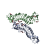
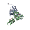
 CHAPERONE / DONOR STRAND COMPLEMENTATION / PILI/N-TERMINAL EXTENSION / PILUS BIOGENESIS / DONOR-STRAND EXCHANGE / NTE / DSC / DSE / PAPA /
  Function and homology information
 Authors
 Citation
 Structure visualization
 Downloads & links
 Other downloads


 PDBj
PDBj
 Assembly
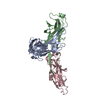
 Mass: 18.015 Da / Num. of mol.: 155 / Source method: isolated from a natural source / Formula: H2O
 Sample preparation
 / Beamline: ID29 / Wavelength: 0.976
  Processing