 Movie
Movie Controller

[English] 日本語
 Yorodumi
- PDB-6zl1: Crystal structure of human serum albumin in complex with the MCL-... -
+ Open data

- Basic information
| Entry | Database: PDB / ID: 6zl1 | ||||||
|---|---|---|---|---|---|---|---|
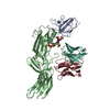
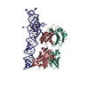
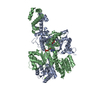
| Title | Crystal structure of human serum albumin in complex with the MCL-1 neutralizing Alphabody CMPX-383B | ||||||
 Components Components |
| ||||||
 Keywords Keywords |  DE NOVO PROTEIN / de novo protein design / 3 helical antiparallel coiled coil / cell penetrating Alphabody MCL-1 neutralizing Alphabody DE NOVO PROTEIN / de novo protein design / 3 helical antiparallel coiled coil / cell penetrating Alphabody MCL-1 neutralizing Alphabody | ||||||
| Function / homology |  Function and homology information Function and homology informationcellular response to calcium ion starvation / exogenous protein binding / Ciprofloxacin ADME / HDL remodeling / enterobactin binding / Heme biosynthesis / negative regulation of mitochondrial depolarization / Prednisone ADME / Heme degradation / antioxidant activity ...cellular response to calcium ion starvation / exogenous protein binding / Ciprofloxacin ADME / HDL remodeling / enterobactin binding / Heme biosynthesis / negative regulation of mitochondrial depolarization / Prednisone ADME / Heme degradation / antioxidant activity / Aspirin ADME / toxic substance binding / Scavenging of heme from plasma / Recycling of bile acids and salts / cellular response to starvation / platelet alpha granule lumen / fatty acid binding / Post-translational protein phosphorylation / Cytoprotection by HMOX1 / Regulation of Insulin-like Growth Factor (IGF) transport and uptake by Insulin-like Growth Factor Binding Proteins (IGFBPs) / pyridoxal phosphate binding / Platelet degranulation / protein-folding chaperone binding / blood microparticle / copper ion binding / endoplasmic reticulum lumen / Golgi apparatus / endoplasmic reticulum / protein-containing complex / DNA binding / extracellular space / extracellular exosome / extracellular region / identical protein binding / nucleus / cytoplasmSimilarity search - Function | ||||||
| Biological species |  Homo sapiens (human) Homo sapiens (human)synthetic construct (others) | ||||||
| Method | X-RAY DIFFRACTION / SYNCHROTRON / MOLECULAR REPLACEMENT / Resolution: 3.272 Å | ||||||
 Authors Authors | Pannecoucke, E. / Savvides, S.N. / Desmet, J. / Lasters, I. | ||||||
| Funding support |  Belgium, 1items Belgium, 1items
| ||||||
 Citation Citation | Journal: Sci Adv / Year: 2021 Title: Cell-penetrating Alphabody protein scaffolds for intracellular drug targeting. Authors: Pannecoucke, E. / Van Trimpont, M. / Desmet, J. / Pieters, T. / Reunes, L. / Demoen, L. / Vuylsteke, M. / Loverix, S. / Vandenbroucke, K. / Alard, P. / Henderikx, P. / Deroo, S. / Baatz, F. ...Authors: Pannecoucke, E. / Van Trimpont, M. / Desmet, J. / Pieters, T. / Reunes, L. / Demoen, L. / Vuylsteke, M. / Loverix, S. / Vandenbroucke, K. / Alard, P. / Henderikx, P. / Deroo, S. / Baatz, F. / Lorent, E. / Thiolloy, S. / Somers, K. / McGrath, Y. / Van Vlierberghe, P. / Lasters, I. / Savvides, S.N. | ||||||
| History |
|
- Structure visualization
| Structure viewer | Molecule: MolmilJmol/JSmol |
|---|
- Downloads & links
-Download
| PDBx/mmCIF format | 6zl1.cif.gz | 468.1 KB | Display | PDBx/mmCIF format |
|---|---|---|---|---|
| PDB format | pdb6zl1.ent.gz | 382.4 KB | Display | PDB format |
| PDBx/mmJSON format | 6zl1.json.gz | Tree view | PDBx/mmJSON format | |
| Others |  Other downloads Other downloads |
-Validation report
| Arichive directory | https://data.pdbj.org/pub/pdb/validation_reports/zl/6zl1ftp://data.pdbj.org/pub/pdb/validation_reports/zl/6zl1 | HTTPS FTP |
|---|
-Related structure data
| Related structure data |  6zieC  3mk8S C: citing same article ( S: Starting model for refinement |
|---|---|
| Similar structure data |


-Links
 PDBj
PDBj


- Assembly
| Deposited unit | 
| ||||||||||
|---|---|---|---|---|---|---|---|---|---|---|---|
| 1 | 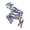
| ||||||||||
| 2 | 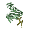
| ||||||||||
| Unit cell |
|
-Components
| #1: Protein | Mass: 69383.719 Da / Num. of mol.: 2 Source method: isolated from a genetically manipulated source Source: (gene. exp.) Homo sapiens (human)Gene: ALB, GIG20, GIG42, PRO0903, PRO1708, PRO2044, PRO2619, PRO2675, UNQ696/PRO1341 Production host: Homo sapiens (human) / References: UniProt: P02768#2: Protein | Mass: 14938.014 Da / Num. of mol.: 2 / Mutation: I92T Source method: isolated from a genetically manipulated source Source: (gene. exp.) synthetic construct (others) / Production host:  Escherichia coli BL21(DE3) (bacteria) Escherichia coli BL21(DE3) (bacteria) |
|---|
-Experimental details
-Experiment
| Experiment | Method: X-RAY DIFFRACTION / Number of used crystals: 1 |
|---|
- Sample preparation
| Crystal | Density Matthews: 2.59 Å3/Da / Density % sol: 52.48 % / Description: Plate-like |
|---|---|
| Crystal grow | Temperature: 310.15 K / Method: vapor diffusion, sitting drop / pH: 6.8 Details: 18% (w/v) PEG 3350, 0.1 M BIS-Tris pH 6.8, 0.2 M ammonium phosphate |
-Data collection
| Diffraction | Mean temperature: 100 K / Serial crystal experiment: N |
|---|---|
| Diffraction source | Source: SYNCHROTRON / Site: PETRA III, EMBL c/o DESY  / Beamline: P14 (MX2) / Wavelength: 0.9763 Å / Beamline: P14 (MX2) / Wavelength: 0.9763 Å |
| Detector | Type: DECTRIS PILATUS 6M-F / Detector: PIXEL / Date: Oct 24, 2016 |
| Radiation | Protocol: SINGLE WAVELENGTH / Monochromatic (M) / Laue (L): M / Scattering type: x-ray |
| Radiation wavelength | Wavelength: 0.9763 Å / Relative weight: 1 |
| Reflection | Resolution: 3.272→115.673 Å / Num. obs: 24556 / % possible obs: 92.1 % / Redundancy: 4.8 % / Biso Wilson estimate: 88.79 Å2 / CC1/2: 0.993 / Net I/σ(I): 8.6 |
| Reflection shell | Resolution: 3.272→3.389 Å / Redundancy: 2.7 % / Mean I/σ(I) obs: 0.52 / Num. unique obs: 1168 / CC1/2: 0.182 / CC star: 0.555 / % possible all: 44.11 |
- Processing
| Software |
| |||||||||||||||||||||||||||||||||||||||||||||||||||||||||||||||||||||||||||||||||||||||||||||||||||||||||||||||||||||||||||||
|---|---|---|---|---|---|---|---|---|---|---|---|---|---|---|---|---|---|---|---|---|---|---|---|---|---|---|---|---|---|---|---|---|---|---|---|---|---|---|---|---|---|---|---|---|---|---|---|---|---|---|---|---|---|---|---|---|---|---|---|---|---|---|---|---|---|---|---|---|---|---|---|---|---|---|---|---|---|---|---|---|---|---|---|---|---|---|---|---|---|---|---|---|---|---|---|---|---|---|---|---|---|---|---|---|---|---|---|---|---|---|---|---|---|---|---|---|---|---|---|---|---|---|---|---|---|---|
| Refinement | Method to determine structure: MOLECULAR REPLACEMENT Starting model: 3MK8 Resolution: 3.272→115.67 Å / Cor.coef. Fo:Fc: 0.898 / Cor.coef. Fo:Fc free: 0.871 / Cross valid method: THROUGHOUT / SU Rfree Blow DPI: 0.556
| |||||||||||||||||||||||||||||||||||||||||||||||||||||||||||||||||||||||||||||||||||||||||||||||||||||||||||||||||||||||||||||
| Displacement parameters | Biso mean: 96.5 Å2
| |||||||||||||||||||||||||||||||||||||||||||||||||||||||||||||||||||||||||||||||||||||||||||||||||||||||||||||||||||||||||||||
| Refine analyze | Luzzati coordinate error obs: 0.55 Å | |||||||||||||||||||||||||||||||||||||||||||||||||||||||||||||||||||||||||||||||||||||||||||||||||||||||||||||||||||||||||||||
| Refinement step | Cycle: LAST / Resolution: 3.272→115.67 Å
| |||||||||||||||||||||||||||||||||||||||||||||||||||||||||||||||||||||||||||||||||||||||||||||||||||||||||||||||||||||||||||||
| Refine LS restraints |
| |||||||||||||||||||||||||||||||||||||||||||||||||||||||||||||||||||||||||||||||||||||||||||||||||||||||||||||||||||||||||||||
| LS refinement shell | Resolution: 3.272→3.34 Å
| |||||||||||||||||||||||||||||||||||||||||||||||||||||||||||||||||||||||||||||||||||||||||||||||||||||||||||||||||||||||||||||
| Refinement TLS params. | Method: refined / Refine-ID: X-RAY DIFFRACTION
| |||||||||||||||||||||||||||||||||||||||||||||||||||||||||||||||||||||||||||||||||||||||||||||||||||||||||||||||||||||||||||||
| Refinement TLS group |
|
