 Movie
Movie Controller

[English] 日本語
 Yorodumi
- PDB-1p58: Complex Organization of Dengue Virus Membrane Proteins as Reveale... -
+ Open data

- Basic information
| Entry | Database: PDB / ID: 1p58 | ||||||
|---|---|---|---|---|---|---|---|
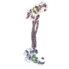
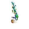
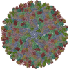
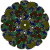
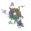
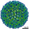
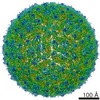
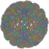
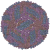
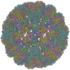
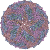
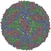
| Title | Complex Organization of Dengue Virus Membrane Proteins as Revealed by 9.5 Angstrom Cryo-EM reconstruction | ||||||
 Components Components |
| ||||||
 Keywords Keywords | VIRUS / FLAVIVIRUS / FLAVIVIRIDAE / DENGUE VIRUS / GLYCOPROTEIN E FROM TICK-BORNE ENCEPHALITIS VIRUS / MEMBRANE PROTEIN M / CRYO-EM / Icosahedral virus | ||||||
| Function / homology |  Function and homology information Function and homology informationsymbiont-mediated suppression of host JAK-STAT cascade via inhibition of host TYK2 activity / flavivirin / host cell mitochondrion / symbiont-mediated suppression of host JAK-STAT cascade via inhibition of STAT2 activity / symbiont-mediated suppression of host cytoplasmic pattern recognition receptor signaling pathway via inhibition of MAVS activity / ribonucleoside triphosphate phosphatase activity / viral capsid / double-stranded RNA binding / nucleoside-triphosphate phosphatase / protein complex oligomerization ...symbiont-mediated suppression of host JAK-STAT cascade via inhibition of host TYK2 activity / flavivirin / host cell mitochondrion / symbiont-mediated suppression of host JAK-STAT cascade via inhibition of STAT2 activity / symbiont-mediated suppression of host cytoplasmic pattern recognition receptor signaling pathway via inhibition of MAVS activity / ribonucleoside triphosphate phosphatase activity / viral capsid / double-stranded RNA binding / nucleoside-triphosphate phosphatase / protein complex oligomerization / monoatomic ion channel activity / mRNA (guanine-N7)-methyltransferase / methyltransferase cap1 / clathrin-dependent endocytosis of virus by host cell / mRNA (nucleoside-2'-O-)-methyltransferase activity / mRNA 5'-cap (guanine-N7-)-methyltransferase activity / RNA helicase activity / host cell perinuclear region of cytoplasm / host cell endoplasmic reticulum membrane / protein dimerization activity / symbiont-mediated suppression of host type I interferon-mediated signaling pathway / RNA helicase / induction by virus of host autophagy / serine-type endopeptidase activity / RNA-directed RNA polymerase / viral RNA genome replication / virus-mediated perturbation of host defense response / RNA-dependent RNA polymerase activity / fusion of virus membrane with host endosome membrane / viral envelope / host cell nucleus / virion attachment to host cell / structural molecule activity / virion membrane / ATP hydrolysis activity / proteolysis / extracellular region / ATP binding / membrane / metal ion binding Similarity search - Function | ||||||
| Biological species |  Dengue virus 2 Puerto Rico/PR159-S1/1969 Dengue virus 2 Puerto Rico/PR159-S1/1969 | ||||||
| Method | ELECTRON MICROSCOPY / single particle reconstruction / cryo EM / Resolution: 9.5 Å | ||||||
 Authors Authors | Zhang, W. / Chipman, P.R. / Corver, J. / Johnson, P.R. / Zhang, Y. / Mukhopadhyay, S. / Baker, T.S. / Strauss, J.H. / Rossmann, M.G. / Kuhn, R.J. | ||||||
 Citation Citation | Journal: Nat Struct Biol / Year: 2003 Title: Visualization of membrane protein domains by cryo-electron microscopy of dengue virus. Authors: Wei Zhang / Paul R Chipman / Jeroen Corver / Peter R Johnson / Ying Zhang / Suchetana Mukhopadhyay / Timothy S Baker / James H Strauss / Michael G Rossmann / Richard J Kuhn /  Abstract: Improved technology for reconstructing cryo-electron microscopy (cryo-EM) images has now made it possible to determine secondary structural features of membrane proteins in enveloped viruses. The ...Improved technology for reconstructing cryo-electron microscopy (cryo-EM) images has now made it possible to determine secondary structural features of membrane proteins in enveloped viruses. The structure of mature dengue virus particles was determined to a resolution of 9.5 A by cryo-EM and image reconstruction techniques, establishing the secondary structural disposition of the 180 envelope (E) and 180 membrane (M) proteins in the lipid envelope. The alpha-helical 'stem' regions of the E molecules, as well as part of the N-terminal section of the M proteins, are buried in the outer leaflet of the viral membrane. The 'anchor' regions of E and the M proteins each form antiparallel E-E and M-M transmembrane alpha-helices, leaving their C termini on the exterior of the viral membrane, consistent with the predicted topology of the unprocessed polyprotein. This is one of only a few determinations of the disposition of transmembrane proteins in situ and shows that the nucleocapsid core and envelope proteins do not have a direct interaction in the mature virus. #1: Journal: Cell(Cambridge,Mass.) / Year: 2002Title: Structure of Dengue Virus: Implications for Flaviviruses Organization, Maturation and Fusion Authors: Kuhn, R.J. / Zhang, W. / Rossmann, M.G. / Pletnev, S.V. / Corver, J. / Lenches, E. / Jones, C.T. / Mukhopadhyay, S. / Chipman, P.R. / Strauss, E.G. / Baker, T.S. / Strauss, J.H. #2: Journal: Nature / Year: 1995Title: The envelope glycoprotein from tick-borne encephalitis virus at 2 A resolution Authors: Rey, F.A. / Heinz, F.X. / Mandl, C. / Kunz, C. / Harrison, S.C. #3: Journal: Nature / Year: 1995Title: Virology. When it's better to lie low. Authors: Kuhn, R.J. / Rossmann, M.G. #4: Journal: Microbiol.Mol.Biol.Rev. / Year: 1999Title: Adding the third dimension to virus life cycles: three-dimensional reconstruction of icosahedral viruses from cryo-electron micrographs Authors: Baker, T.S. / Olson, N.H. / Fuller, S.D. | ||||||
| History |
| ||||||
| Remark 999 | SEQUENCE Only coordinates for CA atoms were submitted. The deposited sequence is based on the E ...SEQUENCE Only coordinates for CA atoms were submitted. The deposited sequence is based on the E protein of dengue 2 virus S1 strain supplied by Hawaii Biotechnology Group, Inc. (Aiea, Hawaii). |
- Structure visualization
| Movie |
Movie viewer |
|---|---|
| Structure viewer | Molecule: MolmilJmol/JSmol |
- Downloads & links
-Download
| PDBx/mmCIF format | 1p58.cif.gz | 64.7 KB | Display | PDBx/mmCIF format |
|---|---|---|---|---|
| PDB format | pdb1p58.ent.gz | 40.7 KB | Display | PDB format |
| PDBx/mmJSON format | 1p58.json.gz | Tree view | PDBx/mmJSON format | |
| Others |  Other downloads Other downloads |
-Validation report
| Summary document | 1p58_validation.pdf.gz | 304.9 KB | Display | wwPDB validaton report |
|---|---|---|---|---|
| Full document | 1p58_full_validation.pdf.gz | 304.8 KB | Display | |
| Data in XML | 1p58_validation.xml.gz | 1.1 KB | Display | |
| Data in CIF | 1p58_validation.cif.gz | 15.3 KB | Display | |
| Arichive directory | https://data.pdbj.org/pub/pdb/validation_reports/p5/1p58ftp://data.pdbj.org/pub/pdb/validation_reports/p5/1p58 | HTTPS FTP |
-Related structure data
| Related structure data | |
|---|---|
| Similar structure data |


-Links
 PDBj
PDBj


- Assembly
| Deposited unit | 
|
|---|---|
| 1 | x 60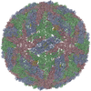
|
| 2 |
|
| 3 | x 5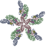
|
| 4 | x 6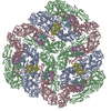
|
| 5 | 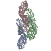
|
| Symmetry | Point symmetry: (Hermann–Mauguin notation: 532 / Schoenflies symbol: I (icosahedral)) |
-Components
| #1: Protein | Mass: 54401.758 Da / Num. of mol.: 3 / Source method: isolated from a natural source / Source: (natural) Dengue virus 2 Puerto Rico/PR159-S1/1969 / Genus: Flavivirus / Species: Dengue virus / Strain: PR159/S1 / References: UniProt: P12823, UniProt: P14337*PLUS#2: Protein | Mass: 8387.841 Da / Num. of mol.: 3 / Source method: isolated from a natural source / Source: (natural) Dengue virus 2 Puerto Rico/PR159-S1/1969 / Genus: Flavivirus / Species: Dengue virus / Strain: PR159/S1 / References: UniProt: P12823, UniProt: Q9WDA7*PLUS |
|---|
-Experimental details
-Experiment
| Experiment | Method: ELECTRON MICROSCOPY |
|---|---|
| EM experiment | Aggregation state: PARTICLE / 3D reconstruction method: single particle reconstruction |
- Sample preparation
| Component | Name: DENGUE VIRUS / Type: VIRUS |
|---|---|
| Details of virus | Host category: INVERTEBRATES / Isolate: STRAIN / Type: VIRION |
| Natural host | Organism: Aedes aegypti / Strain: C6/36 |
| Buffer solution | Name: 50 mM TRIS, 75 mM NaCl, 1 mM EDTA / pH: 7.6 / Details: 50 mM TRIS, 75 mM NaCl, 1 mM EDTA |
| Specimen | Conc.: 20 mg/ml / Embedding applied: NO / Shadowing applied: NO / Staining applied: NO / Vitrification applied: YES |
| Specimen support | Details: Concentration is given in PFU/ML |
| Vitrification | Details: SAMPLES WERE PREPARED AS THIN LAYERS OF VITREOUS ICE AND MAINTAINED AT LIQUID NITROGEN TEMPERATURE IN THE ELECTRON MICROSCOPE |
| Crystal grow | *PLUS Method: electron microscopy / Details: Electron Microscopy |
- Electron microscopy imaging
| Microscopy | Model: FEI/PHILIPS CM200T / Date: Jun 27, 2000 |
|---|---|
| Electron gun | Electron source:  FIELD EMISSION GUN / Accelerating voltage: 200 kV / Illumination mode: FLOOD BEAM FIELD EMISSION GUN / Accelerating voltage: 200 kV / Illumination mode: FLOOD BEAM |
| Electron lens | Mode: BRIGHT FIELD / Nominal magnification: 50000 X / Nominal defocus max: 4800 nm / Nominal defocus min: 800 nm / Cs: 2 mm |
| Specimen holder | Temperature: 87 K / Tilt angle max: 0 ° / Tilt angle min: 0 ° |
| Image recording | Electron dose: 27 e/Å2 / Film or detector model: KODAK SO-163 FILM |
- Processing
| EM software |
| |||||||||||||||||||||
|---|---|---|---|---|---|---|---|---|---|---|---|---|---|---|---|---|---|---|---|---|---|---|
| CTF correction | Details: each viral image was CTF corrected before reconstruction, based on the following equation: F(corr)=F(obs)/[|CTF|+wiener*(1-|CTF|)] | |||||||||||||||||||||
| Symmetry | Point symmetry: I (icosahedral) | |||||||||||||||||||||
| 3D reconstruction | Method: FOURIER-BESSEL METHOD / Resolution: 9.5 Å / Num. of particles: 1691 / Nominal pixel size: 2.8 Å Details: THE RECONSTRUCTION WAS COMPUTED FROM 1691 DENGUE VIRUS IMAGES THAT WERE SELECTED FROM 78 MICROGRAPHS. ORIENTATIONS WERE DETERMINED BY THE MODEL-BASED POLAR-FOURIER TRANSFORM METHOD (BAKER ...Details: THE RECONSTRUCTION WAS COMPUTED FROM 1691 DENGUE VIRUS IMAGES THAT WERE SELECTED FROM 78 MICROGRAPHS. ORIENTATIONS WERE DETERMINED BY THE MODEL-BASED POLAR-FOURIER TRANSFORM METHOD (BAKER AND CHENG, 1996, J.STRUC.BIOL. 116,120-130) AND REFINED BY THE MODEL-BASED FOURIER TRANSFORM REFINEMENT PROCEDURE(http://bond.cs.ucf.edu/ComputationalBiology/Projects/POR/Home.html). Symmetry type: POINT | |||||||||||||||||||||
| Atomic model building | Space: REAL | |||||||||||||||||||||
| Atomic model building |
| |||||||||||||||||||||
| Refinement step | Cycle: LAST
|
