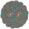
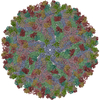
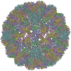
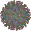
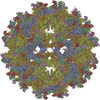
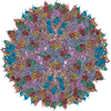
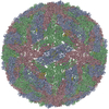
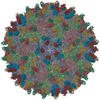
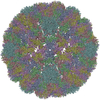
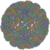
CryoEM Structure of Merkel Cell Polyomavirus Virus-like Particle
Components
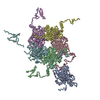
Capsid protein VP1
Keywords
VIRUS LIKE PARTICLE / polyomavirus / capsid / Merkel cell carcinoma / jelly roll fold
Function / homology
Capsid protein VP1,Polyomavirus / Polyomavirus capsid protein VP1 superfamily / Polyomavirus coat protein / Double-stranded DNA virus, group I, capsid / viral capsid / structural molecule activity / Capsid protein VP1
Function and homology information
Biological species
Merkel cell polyomavirus
Method
ELECTRON MICROSCOPY / single particle reconstruction / cryo EM / Resolution: 3.4 Å
Journal: J Virol / Year: 2020 Title: Structure of Merkel Cell Polyomavirus Capsid and Interaction with Its Glycosaminoglycan Attachment Receptor. Authors: Niklas J Bayer / Dovile Januliene / Georg Zocher / Thilo Stehle / Arne Moeller / Bärbel S Blaum / Abstract: Merkel cell polyomavirus (MCPyV) is a human double-stranded DNA tumor virus. MCPyV cell entry is unique among members of the polyomavirus family as it requires the engagement of two types of glycans, ...Merkel cell polyomavirus (MCPyV) is a human double-stranded DNA tumor virus. MCPyV cell entry is unique among members of the polyomavirus family as it requires the engagement of two types of glycans, sialylated oligosaccharides and sulfated glycosaminoglycans (GAGs). Here, we present crystallographic and cryo-electron microscopic structures of the icosahedral MCPyV capsid and analysis of its glycan interactions via nuclear magnetic resonance (NMR) spectroscopy. While sialic acid binding is specific for α2-3-linked sialic acid and mediated by the exposed apical loops of the major capsid protein VP1, a broad range of GAG oligosaccharides bind to recessed regions between VP1 capsomers. Individual VP1 capsomers are tethered to one another by an extensive disulfide network that differs in architecture from previously described interactions for other PyVs. An unusual C-terminal extension in MCPyV VP1 projects from the recessed capsid regions. Mutagenesis experiments show that this extension is dispensable for receptor interactions. The MCPyV genome was found to be clonally integrated in 80% of cases of Merkel cell carcinoma (MCC), a rare but aggressive form of human skin cancer, strongly suggesting that this virus is tumorigenic. In the metastasizing state, the course of the disease is often fatal, especially in immunocompromised individuals, as reflected by the high mortality rate of 33 to 46% and the low 5-year survival rate (<45%). The high seroprevalence of about 60% makes MCPyV a serious health care burden and illustrates the need for targeted treatments. In this study, we present the first high-resolution structural data for this human tumor virus and demonstrate that the full capsid is required for the essential interaction with its GAG receptor(s). Together, these data can be used as a basis for future strategies in drug development.
Average exposure time: 67 sec. / Electron dose: 30 e/Å2 / Detector mode: COUNTING / Film or detector model: FEI FALCON III (4k x 4k) / Num. of grids imaged: 1 / Num. of real images: 463
-
Processing
EM software
ID
Name
Version
Category
1
DoG Picker
particleselection
2
EPU
imageacquisition
4
CTFFIND
4
CTFcorrection
7
UCSF Chimera
1.11.2
modelfitting
9
PHENIX
1.14
modelrefinement
10
Coot
0.8.9.1
modelrefinement
11
FREALIGN
initialEulerassignment
12
cisTEM
finalEulerassignment
13
cisTEM
classification
14
cisTEM
3Dreconstruction
CTF correction
Details: CTF correction was performed within 3D reconstruction Type: NONE
Particle selection
Num. of particles selected: 23349
Symmetry
Point symmetry: I (icosahedral)
3D reconstruction
Resolution: 3.4 Å / Resolution method: FSC 0.143 CUT-OFF / Num. of particles: 22310 / Num. of class averages: 1 / Symmetry type: POINT
Atomic model building
B value: 105.92 / Protocol: FLEXIBLE FIT / Space: REAL / Target criteria: Correlation coefficient
In the structure databanks used in Yorodumi, some data are registered as the other names, "COVID-19 virus" and "2019-nCoV". Here are the details of the virus and the list of structure data.
Jan 31, 2019. EMDB accession codes are about to change! (news from PDBe EMDB page)
EMDB accession codes are about to change! (news from PDBe EMDB page)
The allocation of 4 digits for EMDB accession codes will soon come to an end. Whilst these codes will remain in use, new EMDB accession codes will include an additional digit and will expand incrementally as the available range of codes is exhausted. The current 4-digit format prefixed with “EMD-” (i.e. EMD-XXXX) will advance to a 5-digit format (i.e. EMD-XXXXX), and so on. It is currently estimated that the 4-digit codes will be depleted around Spring 2019, at which point the 5-digit format will come into force.
The EM Navigator/Yorodumi systems omit the EMD- prefix.
Related info.:Q: What is EMD? / ID/Accession-code notation in Yorodumi/EM Navigator
Yorodumi is a browser for structure data from EMDB, PDB, SASBDB, etc.
This page is also the successor to EM Navigator detail page, and also detail information page/front-end page for Omokage search.
The word "yorodu" (or yorozu) is an old Japanese word meaning "ten thousand". "mi" (miru) is to see.
Related info.:EMDB / PDB / SASBDB / Comparison of 3 databanks / Yorodumi Search / Aug 31, 2016. New EM Navigator & Yorodumi / Yorodumi Papers / Jmol/JSmol / Function and homology information / Changes in new EM Navigator and Yorodumi
 Movie
Movie Controller


 Open data
 Basic information
 Components
 Keywords
 Function and homology information
 Authors
 Germany, 3items
  Citation
 Structure visualization
 Downloads & links
 Other downloads
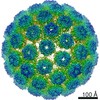

 PDBj
PDBj


 Assembly

 Sample preparation
 Electron microscopy imaging
 Processing