 Movie
Movie Controller

[English] 日本語
 Yorodumi
- PDB-6zlz: Crystal Structure of Merkel Cell Polyomavirus Virus-like Particle -
+ Open data

- Basic information
| Entry | Database: PDB / ID: 6zlz | ||||||||||||
|---|---|---|---|---|---|---|---|---|---|---|---|---|---|
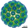
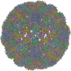
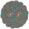
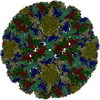
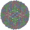
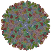
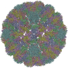
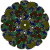
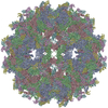
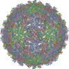
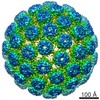
| Title | Crystal Structure of Merkel Cell Polyomavirus Virus-like Particle | ||||||||||||
 Components Components | Capsid protein VP1 | ||||||||||||
 Keywords Keywords | VIRUS LIKE PARTICLE / polyomavirus / capsid / Merkel cell carcinoma / jelly roll fold | ||||||||||||
| Function / homology | Capsid protein VP1,Polyomavirus / Polyomavirus capsid protein VP1 superfamily / Polyomavirus coat protein / Double-stranded DNA virus, group I, capsid / viral capsid / structural molecule activity / Capsid protein VP1 Function and homology information Function and homology information | ||||||||||||
| Biological species |  Merkel cell polyomavirus Merkel cell polyomavirus | ||||||||||||
| Method | X-RAY DIFFRACTION / SYNCHROTRON / MOLECULAR REPLACEMENT / Resolution: 3.52 Å | ||||||||||||
 Authors Authors | Bayer, N.J. / Stehle, T. / Blaum, B.S. | ||||||||||||
| Funding support |  Germany, 3items Germany, 3items
| ||||||||||||
 Citation Citation | Journal: J Virol / Year: 2020 Title: Structure of Merkel Cell Polyomavirus Capsid and Interaction with Its Glycosaminoglycan Attachment Receptor. Authors: Niklas J Bayer / Dovile Januliene / Georg Zocher / Thilo Stehle / Arne Moeller / Bärbel S Blaum /  Abstract: Merkel cell polyomavirus (MCPyV) is a human double-stranded DNA tumor virus. MCPyV cell entry is unique among members of the polyomavirus family as it requires the engagement of two types of glycans, ...Merkel cell polyomavirus (MCPyV) is a human double-stranded DNA tumor virus. MCPyV cell entry is unique among members of the polyomavirus family as it requires the engagement of two types of glycans, sialylated oligosaccharides and sulfated glycosaminoglycans (GAGs). Here, we present crystallographic and cryo-electron microscopic structures of the icosahedral MCPyV capsid and analysis of its glycan interactions via nuclear magnetic resonance (NMR) spectroscopy. While sialic acid binding is specific for α2-3-linked sialic acid and mediated by the exposed apical loops of the major capsid protein VP1, a broad range of GAG oligosaccharides bind to recessed regions between VP1 capsomers. Individual VP1 capsomers are tethered to one another by an extensive disulfide network that differs in architecture from previously described interactions for other PyVs. An unusual C-terminal extension in MCPyV VP1 projects from the recessed capsid regions. Mutagenesis experiments show that this extension is dispensable for receptor interactions. The MCPyV genome was found to be clonally integrated in 80% of cases of Merkel cell carcinoma (MCC), a rare but aggressive form of human skin cancer, strongly suggesting that this virus is tumorigenic. In the metastasizing state, the course of the disease is often fatal, especially in immunocompromised individuals, as reflected by the high mortality rate of 33 to 46% and the low 5-year survival rate (<45%). The high seroprevalence of about 60% makes MCPyV a serious health care burden and illustrates the need for targeted treatments. In this study, we present the first high-resolution structural data for this human tumor virus and demonstrate that the full capsid is required for the essential interaction with its GAG receptor(s). Together, these data can be used as a basis for future strategies in drug development. | ||||||||||||
| History |
|
- Structure visualization
| Structure viewer | Molecule: MolmilJmol/JSmol |
|---|
- Downloads & links
-Download
| PDBx/mmCIF format | 6zlz.cif.gz | 377.1 KB | Display | PDBx/mmCIF format |
|---|---|---|---|---|
| PDB format | pdb6zlz.ent.gz | 312.1 KB | Display | PDB format |
| PDBx/mmJSON format | 6zlz.json.gz | Tree view | PDBx/mmJSON format | |
| Others |  Other downloads Other downloads |
-Validation report
| Arichive directory | https://data.pdbj.org/pub/pdb/validation_reports/zl/6zlzftp://data.pdbj.org/pub/pdb/validation_reports/zl/6zlz | HTTPS FTP |
|---|
-Related structure data
| Related structure data |  6zmlC  1svaS S: Starting model for refinement C: citing same article ( |
|---|---|
| Similar structure data |
-Links
 PDBj
PDBj


- Assembly
| Deposited unit | 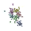
| ||||||||||||||||||||||||||||||||||||||||||||||||||||||||||||||||
|---|---|---|---|---|---|---|---|---|---|---|---|---|---|---|---|---|---|---|---|---|---|---|---|---|---|---|---|---|---|---|---|---|---|---|---|---|---|---|---|---|---|---|---|---|---|---|---|---|---|---|---|---|---|---|---|---|---|---|---|---|---|---|---|---|---|
| 1 | x 60
| ||||||||||||||||||||||||||||||||||||||||||||||||||||||||||||||||
| Unit cell |
| ||||||||||||||||||||||||||||||||||||||||||||||||||||||||||||||||
| Noncrystallographic symmetry (NCS) | NCS oper:
|
-Components
| #1: Protein | / VP1 Mass: 46611.031 Da / Num. of mol.: 6 Source method: isolated from a genetically manipulated source Source: (gene. exp.) Merkel cell polyomavirus / Gene: VP1 / Plasmid: pwM / Cell line (production host): HEK293TT / Organ (production host): Kidney / Production host:  Homo sapiens (human) / References: UniProt: B0G0W3 Homo sapiens (human) / References: UniProt: B0G0W3#2: Chemical | ChemComp-CA /   Mass: 40.078 Da / Num. of mol.: 6 / Source method: obtained synthetically / Formula: Ca / Feature type: SUBJECT OF INVESTIGATION Mass: 40.078 Da / Num. of mol.: 6 / Source method: obtained synthetically / Formula: Ca / Feature type: SUBJECT OF INVESTIGATIONHas ligand of interest | Y | |
|---|
-Experimental details
-Experiment
| Experiment | Method: X-RAY DIFFRACTION / Number of used crystals: 1 |
|---|
- Sample preparation
| Crystal |
| |||
|---|---|---|---|---|
| Crystal grow | Temperature: 293 K / Method: vapor diffusion, hanging drop / pH: 4.6 Details: 0.1 M sodium citrate, 0.2 M sodium chloride, 30% MPD |
-Data collection
| Diffraction | Mean temperature: 100 K / Serial crystal experiment: N |
|---|---|
| Diffraction source | Source: SYNCHROTRON / Site: Diamond  / Beamline: I03 / Wavelength: 0.976 Å / Beamline: I03 / Wavelength: 0.976 Å |
| Detector | Type: DECTRIS PILATUS 6M / Detector: PIXEL / Date: May 25, 2016 |
| Radiation | Monochromator: M / Protocol: SINGLE WAVELENGTH / Monochromatic (M) / Laue (L): M / Scattering type: x-ray |
| Radiation wavelength | Wavelength: 0.976 Å / Relative weight: 1 |
| Reflection | Resolution: 3.52→50 Å / Num. obs: 1088665 / % possible obs: 99.8 % / Redundancy: 13.5 % / Biso Wilson estimate: 97.6 Å2 / CC1/2: 0.996 / Rrim(I) all: 0.309 / Net I/σ(I): 9.15 |
| Reflection shell | Resolution: 3.52→3.6 Å / Redundancy: 12.6 % / Mean I/σ(I) obs: 1.06 / Num. unique obs: 78618 / CC1/2: 0.332 / Rrim(I) all: 2.883 / % possible all: 98.3 |
- Processing
| Software |
| ||||||||||||||||||
|---|---|---|---|---|---|---|---|---|---|---|---|---|---|---|---|---|---|---|---|
| Refinement | Method to determine structure: MOLECULAR REPLACEMENT Starting model: 1sva Resolution: 3.52→49.97 Å / Cross valid method: NONE Details: NCS-constraint refinement procedure with NCS map averaging
| ||||||||||||||||||
| Displacement parameters | Biso max: 190.55 Å2 / Biso mean: 122.6 Å2 / Biso min: 88.69 Å2 | ||||||||||||||||||
| Refinement step | Cycle: LAST / Resolution: 3.52→49.97 Å
|
