 Movie
Movie Controller

[English] 日本語
 Yorodumi
- PDB-1jch: Crystal Structure of Colicin E3 in Complex with its Immunity Protein -
+ Open data

- Basic information
| Entry | Database: PDB / ID: 1jch | ||||||
|---|---|---|---|---|---|---|---|
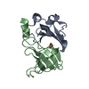
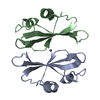
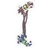
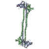
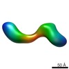
| Title | Crystal Structure of Colicin E3 in Complex with its Immunity Protein | ||||||
 Components Components |
| ||||||
 Keywords Keywords | RIBOSOME INHIBITOR /  HYDROLASE / TRANSLOCATION DOMAIN IS A BETA-JELLYROLL / THE RECEPTOR-BINDING DOMAIN IS A COILED COIL / THE RNASE DOMAIN IS A SIX-STRANDED ANTIPARALLEL BETA-SHEET. THE IMMUNITY PROTEIN IS A FOUR-STRANDED ANTIPARALLEL BETA SHEET FLANKED BY 3 HELICES ON ONE SIDE OF THE SHEET HYDROLASE / TRANSLOCATION DOMAIN IS A BETA-JELLYROLL / THE RECEPTOR-BINDING DOMAIN IS A COILED COIL / THE RNASE DOMAIN IS A SIX-STRANDED ANTIPARALLEL BETA-SHEET. THE IMMUNITY PROTEIN IS A FOUR-STRANDED ANTIPARALLEL BETA SHEET FLANKED BY 3 HELICES ON ONE SIDE OF THE SHEET | ||||||
| Function / homology |  Function and homology information Function and homology informationnegative regulation of ion transmembrane transporter activity / extrachromosomal circular DNA / bacteriocin immunity / toxic substance binding / ribosome binding / Lyases; Phosphorus-oxygen lyases / endonuclease activity / killing of cells of another organism / transmembrane transporter binding / defense response to bacterium / RNA bindingSimilarity search - Function | ||||||
| Biological species |  Escherichia coli str. K12 substr. (bacteria) Escherichia coli str. K12 substr. (bacteria) | ||||||
| Method | X-RAY DIFFRACTION / SYNCHROTRON / MIR / Resolution: 3.02 Å | ||||||
 Authors Authors | Soelaiman, S. / Jakes, K. / Wu, N. / Li, C. / Shoham, M. | ||||||
 Citation Citation | Journal: Mol.Cell / Year: 2001 Title: Crystal structure of colicin E3: implications for cell entry and ribosome inactivation. Authors: Soelaiman, S. / Jakes, K. / Wu, N. / Li, C. / Shoham, M. | ||||||
| History |
| ||||||
| Remark 300 | BIOMOLECULE: 1, 2 THIS ENTRY CONTAINS THE CRYSTALLOGRAPHIC ASYMMETRIC UNIT WHICH CONSISTS OF 2 ... BIOMOLECULE: 1, 2 THIS ENTRY CONTAINS THE CRYSTALLOGRAPHIC ASYMMETRIC UNIT WHICH CONSISTS OF 2 BIOLOGICAL UNITS. THE FIRST BIOLOGICAL UNIT CONTAINS PROTEIN CHAINS A+B AND HETGROUPS CIT 601 AND 602, GOL 701 AND 702 AND HOH RESIDUES NOT STARTING WITH A PREFIX 5000. THE SECOND BIOLOGICAL UNIT CONTAINS PROTEIN CHAINS C+D AND HETGROUPS CIT 5601 AND 5602, GOL 5701 AND 5702 AND HOH RESIDUES STARTING WITH A PREFIX 5000. |
- Structure visualization
| Structure viewer | Molecule: MolmilJmol/JSmol |
|---|
- Downloads & links
-Download
| PDBx/mmCIF format | 1jch.cif.gz | 238.5 KB | Display | PDBx/mmCIF format |
|---|---|---|---|---|
| PDB format | pdb1jch.ent.gz | 189.1 KB | Display | PDB format |
| PDBx/mmJSON format | 1jch.json.gz | Tree view | PDBx/mmJSON format | |
| Others |  Other downloads Other downloads |
-Validation report
| Arichive directory | https://data.pdbj.org/pub/pdb/validation_reports/jc/1jchftp://data.pdbj.org/pub/pdb/validation_reports/jc/1jch | HTTPS FTP |
|---|
-Related structure data
| Related structure data | |
|---|---|
| Similar structure data |


-Links
 PDBj
PDBj- Assembly
| Deposited unit | 
| ||||||||||||
|---|---|---|---|---|---|---|---|---|---|---|---|---|---|
| 1 |
| ||||||||||||
| 2 | 
| ||||||||||||
| 3 | 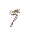
| ||||||||||||
| Unit cell |
| ||||||||||||
| Noncrystallographic symmetry (NCS) | NCS oper:
| ||||||||||||
| Details | COLICIN E3 FORMS A DIMER IN SOLUTION AS WELL AS IN THE CRYSTALLINE STATE. THE SECOND MOLECULE IN THE ASYMMETRIC UNIT CAN BE GENERATED BY THE FOLLOWING MATRIX: -0.99997 0.00024 0.00756, -0.00024 -1.00000 -0.00042, 0.00756 -0.00042 0.99997, AND TRANSLATION VECTOR IN ANGSTROMS: 33.345 148.822 -0.009 |
-Components
| #1: Protein | Mass: 58043.008 Da / Num. of mol.: 2 Source method: isolated from a genetically manipulated source Source: (gene. exp.) Escherichia coli str. K12 substr. (bacteria)Species: Escherichia coli / Strain: W3110 / Species (production host): Escherichia coliProduction host: Escherichia coli str. K12 substr. W3110 (bacteria)Strain (production host): W3110 References: UniProt: P00646, Hydrolases; Acting on ester bonds; Endodeoxyribonucleases producing 5'-phosphomonoesters#2: Protein | Mass: 9779.565 Da / Num. of mol.: 2 Source method: isolated from a genetically manipulated source Source: (gene. exp.) Escherichia coli str. K12 substr. (bacteria)Species: Escherichia coli / Strain: W3110 / Species (production host): Escherichia coliProduction host: Escherichia coli str. K12 substr. W3110 (bacteria)Strain (production host): W3110 / References: UniProt: P02984 #3: Chemical | ChemComp-CIT / Citric acid  Mass: 192.124 Da / Num. of mol.: 4 / Source method: obtained synthetically / Formula: C6H8O7 Mass: 192.124 Da / Num. of mol.: 4 / Source method: obtained synthetically / Formula: C6H8O7#4: Chemical | ChemComp-GOL / Glycerol  Mass: 92.094 Da / Num. of mol.: 4 / Source method: obtained synthetically / Formula: C3H8O3 Mass: 92.094 Da / Num. of mol.: 4 / Source method: obtained synthetically / Formula: C3H8O3#5: Water | ChemComp-HOH / | Water Mass: 18.015 Da / Num. of mol.: 396 / Source method: isolated from a natural source / Formula: H2O Mass: 18.015 Da / Num. of mol.: 396 / Source method: isolated from a natural source / Formula: H2O |
|---|
-Experimental details
-Experiment
| Experiment | Method: X-RAY DIFFRACTION / Number of used crystals: 1 |
|---|
- Sample preparation
| Crystal | Density Matthews: 3.62 Å3/Da / Density % sol: 66 % |
|---|---|
| Crystal grow | Temperature: 277 K / Method: vapor diffusion, hanging drop / pH: 5.6 Details: SODIUM CITRATE, CADMIUM ACETATE, ph 5.6, VAPOR DIFFUSION, HANGING DROP at 277K |
| Crystal grow | *PLUS Temperature: 4 ℃ |
| Components of the solutions | *PLUS Conc.: 1 M / Common name: sodium citrate / Details: pH5.6 |
-Data collection
| Diffraction | Mean temperature: 100 K |
|---|---|
| Diffraction source | Source: SYNCHROTRON / Site: APS  / Beamline: 14-BM-C / Wavelength: 1.037 Å / Beamline: 14-BM-C / Wavelength: 1.037 Å |
| Detector | Type: ADSC QUANTUM 4 / Detector: CCD / Date: Nov 19, 1998 |
| Radiation | Monochromator: GRAPHITE / Protocol: SINGLE WAVELENGTH / Monochromatic (M) / Laue (L): M / Scattering type: x-ray |
| Radiation wavelength | Wavelength: 1.037 Å / Relative weight: 1 |
| Reflection | Resolution: 3.02→20 Å / Num. all: 37014 / Num. obs: 37014 / % possible obs: 94.1 % / Observed criterion σ(I): -3 / Redundancy: 3.2 % / Biso Wilson estimate: 70.8 Å2 / Rmerge(I) obs: 0.067 / Net I/σ(I): 19.5 |
| Reflection shell | Resolution: 3→3.11 Å / Redundancy: 3.2 % / Rmerge(I) obs: 0.197 / Mean I/σ(I) obs: 3.8 / Num. unique all: 3509 / % possible all: 86.4 |
- Processing
| Software |
| ||||||||||||||||||||||||||||||||||||
|---|---|---|---|---|---|---|---|---|---|---|---|---|---|---|---|---|---|---|---|---|---|---|---|---|---|---|---|---|---|---|---|---|---|---|---|---|---|
| Refinement | Method to determine structure: MIR / Resolution: 3.02→20.17 Å / Rfactor Rfree error: 0.008 / Data cutoff high absF: 2270567.39 / Data cutoff low absF: 0 / Isotropic thermal model: RESTRAINED / Cross valid method: THROUGHOUT / σ(F): 0 / σ(I): 0 / Stereochemistry target values: Engh & Huber
| ||||||||||||||||||||||||||||||||||||
| Solvent computation | Solvent model: FLAT MODEL / Bsol: 64.5511 Å2 / ksol: 0.26419 e/Å3 | ||||||||||||||||||||||||||||||||||||
| Displacement parameters | Biso mean: 74.7 Å2
| ||||||||||||||||||||||||||||||||||||
| Refine analyze |
| ||||||||||||||||||||||||||||||||||||
| Refinement step | Cycle: LAST / Resolution: 3.02→20.17 Å
| ||||||||||||||||||||||||||||||||||||
| Refine LS restraints |
| ||||||||||||||||||||||||||||||||||||
| LS refinement shell | Resolution: 3→3.14 Å / Total num. of bins used: 8
| ||||||||||||||||||||||||||||||||||||
| Xplor file |
| ||||||||||||||||||||||||||||||||||||
| Software | *PLUS Name: CNS / Version: 1 / Classification: refinement | ||||||||||||||||||||||||||||||||||||
| Refinement | *PLUS σ(F): 0 / % reflection Rfree: 3 % / Rfactor obs: 0.233 / Rfactor Rfree: 0.282 | ||||||||||||||||||||||||||||||||||||
| Solvent computation | *PLUS | ||||||||||||||||||||||||||||||||||||
| Displacement parameters | *PLUS Biso mean: 74.7 Å2 | ||||||||||||||||||||||||||||||||||||
| Refine LS restraints | *PLUS
| ||||||||||||||||||||||||||||||||||||
| LS refinement shell | *PLUS Rfactor Rfree: 0.297 / % reflection Rfree: 2.6 % / Rfactor Rwork: 0.323 |
