 Movie
Movie Controller

[English] 日本語
 Yorodumi
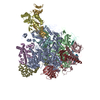
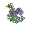
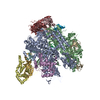
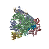
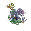
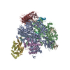
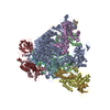
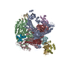
- PDB-3j0k: Orientation of RNA polymerase II within the human VP16-Mediator-p... -
+ Open data

- Basic information
| Entry | Database: PDB / ID: 3j0k | ||||||
|---|---|---|---|---|---|---|---|
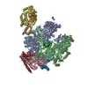
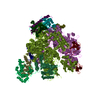
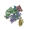
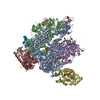
| Title | Orientation of RNA polymerase II within the human VP16-Mediator-pol II-TFIIF assembly | ||||||
 Components Components |
| ||||||
 Keywords Keywords | TRANSFERASE/TRANSCRIPTION / TRANSFERASE-TRANSCRIPTION complex | ||||||
| Function / homology |  Function and homology information Function and homology informationRPB4-RPB7 complex / nuclear-transcribed mRNA catabolic process, deadenylation-dependent decay / RNA Polymerase I Transcription Initiation / Processing of Capped Intron-Containing Pre-mRNA / RNA Polymerase III Transcription Initiation From Type 2 Promoter / RNA Pol II CTD phosphorylation and interaction with CE / Formation of the Early Elongation Complex / mRNA Capping / RNA polymerase II transcribes snRNA genes / TP53 Regulates Transcription of DNA Repair Genes ...RPB4-RPB7 complex / nuclear-transcribed mRNA catabolic process, deadenylation-dependent decay / RNA Polymerase I Transcription Initiation / Processing of Capped Intron-Containing Pre-mRNA / RNA Polymerase III Transcription Initiation From Type 2 Promoter / RNA Pol II CTD phosphorylation and interaction with CE / Formation of the Early Elongation Complex / mRNA Capping / RNA polymerase II transcribes snRNA genes / TP53 Regulates Transcription of DNA Repair Genes / termination of RNA polymerase II transcription / RNA Polymerase II Promoter Escape / RNA Polymerase II Transcription Pre-Initiation And Promoter Opening / RNA Polymerase II Transcription Initiation / RNA Polymerase II Transcription Initiation And Promoter Clearance / RNA-templated transcription / RNA Polymerase II Pre-transcription Events / termination of RNA polymerase III transcription / Formation of TC-NER Pre-Incision Complex / termination of RNA polymerase I transcription / transcription initiation at RNA polymerase III promoter / RNA Polymerase I Promoter Escape / nucleolar large rRNA transcription by RNA polymerase I / maintenance of transcriptional fidelity during transcription elongation by RNA polymerase II / Gap-filling DNA repair synthesis and ligation in TC-NER / transcription initiation at RNA polymerase I promoter / transcription by RNA polymerase I / Estrogen-dependent gene expression / positive regulation of nuclear-transcribed mRNA poly(A) tail shortening / nuclear-transcribed mRNA catabolic process / transcription by RNA polymerase III / RNA polymerase II activity / Dual incision in TC-NER / transcription elongation by RNA polymerase I / tRNA transcription by RNA polymerase III / RNA polymerase I activity / RNA polymerase I complex / RNA polymerase III complex / positive regulation of translational initiation / transcription-coupled nucleotide-excision repair / RNA polymerase II, core complex / translesion synthesis / translation initiation factor binding / DNA-templated transcription initiation / transcription initiation at RNA polymerase II promoter / transcription elongation by RNA polymerase II / P-body / ribonucleoside binding / DNA-directed 5'-3' RNA polymerase activity / DNA-directed RNA polymerase / cytoplasmic stress granule / mRNA processing / ribosome biogenesis / single-stranded DNA binding / nucleic acid binding / transcription by RNA polymerase II / single-stranded RNA binding / protein dimerization activity / mRNA binding / nucleotide binding / nucleolus / mitochondrion / DNA binding / zinc ion binding / nucleoplasm / nucleus / metal ion binding / cytoplasm Similarity search - Function | ||||||
| Biological species |  Homo sapiens (human) Homo sapiens (human) | ||||||
| Method | ELECTRON MICROSCOPY / single particle reconstruction / cryo EM / Resolution: 36 Å | ||||||
 Authors Authors | Bernecky, C. / Grob, P. / Ebmeier, C.C. / Nogales, E. / Taatjes, D.J. | ||||||
 Citation Citation | Journal: PLoS Biol / Year: 2011 Title: Molecular architecture of the human Mediator-RNA polymerase II-TFIIF assembly. Authors: Carrie Bernecky / Patricia Grob / Christopher C Ebmeier / Eva Nogales / Dylan J Taatjes /  Abstract: The macromolecular assembly required to initiate transcription of protein-coding genes, known as the Pre-Initiation Complex (PIC), consists of multiple protein complexes and is approximately 3.5 MDa ...The macromolecular assembly required to initiate transcription of protein-coding genes, known as the Pre-Initiation Complex (PIC), consists of multiple protein complexes and is approximately 3.5 MDa in size. At the heart of this assembly is the Mediator complex, which helps regulate PIC activity and interacts with the RNA polymerase II (pol II) enzyme. The structure of the human Mediator-pol II interface is not well-characterized, whereas attempts to structurally define the Mediator-pol II interaction in yeast have relied on incomplete assemblies of Mediator and/or pol II and have yielded inconsistent interpretations. We have assembled the complete, 1.9 MDa human Mediator-pol II-TFIIF complex from purified components and have characterized its structural organization using cryo-electron microscopy and single-particle reconstruction techniques. The orientation of pol II within this assembly was determined by crystal structure docking and further validated with projection matching experiments, allowing the structural organization of the entire human PIC to be envisioned. Significantly, pol II orientation within the Mediator-pol II-TFIIF assembly can be reconciled with past studies that determined the location of other PIC components relative to pol II itself. Pol II surfaces required for interacting with TFIIB, TFIIE, and promoter DNA (i.e., the pol II cleft) are exposed within the Mediator-pol II-TFIIF structure; RNA exit is unhindered along the RPB4/7 subunits; upstream and downstream DNA is accessible for binding additional factors; and no major structural re-organization is necessary to accommodate the large, multi-subunit TFIIH or TFIID complexes. The data also reveal how pol II binding excludes Mediator-CDK8 subcomplex interactions and provide a structural basis for Mediator-dependent control of PIC assembly and function. Finally, parallel structural analysis of Mediator-pol II complexes lacking TFIIF reveal that TFIIF plays a key role in stabilizing pol II orientation within the assembly. | ||||||
| History |
|
- Structure visualization
| Movie |
Movie viewer |
|---|---|
| Structure viewer | Molecule: MolmilJmol/JSmol |
- Downloads & links
-Download
| PDBx/mmCIF format | 3j0k.cif.gz | 789.7 KB | Display | PDBx/mmCIF format |
|---|---|---|---|---|
| PDB format | pdb3j0k.ent.gz | 641.6 KB | Display | PDB format |
| PDBx/mmJSON format | 3j0k.json.gz | Tree view | PDBx/mmJSON format | |
| Others |  Other downloads Other downloads |
-Validation report
| Summary document | 3j0k_validation.pdf.gz | 842 KB | Display | wwPDB validaton report |
|---|---|---|---|---|
| Full document | 3j0k_full_validation.pdf.gz | 1.2 MB | Display | |
| Data in XML | 3j0k_validation.xml.gz | 142.5 KB | Display | |
| Data in CIF | 3j0k_validation.cif.gz | 212.4 KB | Display | |
| Arichive directory | https://data.pdbj.org/pub/pdb/validation_reports/j0/3j0kftp://data.pdbj.org/pub/pdb/validation_reports/j0/3j0k | HTTPS FTP |
-Related structure data
| Related structure data | 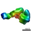 5343MC M: map data used to model this data C: citing same article ( |
|---|---|
| Similar structure data |


-Links
 PDBj
PDBj


- Assembly
| Deposited unit | 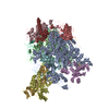
|
|---|---|
| 1 |
|
-Components
-DNA-directed RNA polymerase II ... , 7 types, 7 molecules ABCDGIK
| #1: Protein | Mass: 163180.016 Da / Num. of mol.: 1 / Source method: isolated from a natural source / Source: (natural) Homo sapiens (human) / Cell line: HeLa / Organelle: nucleusReferences: UniProt: P04050*PLUS, DNA-directed RNA polymerase |
|---|---|
| #2: Protein | Mass: 138937.297 Da / Num. of mol.: 1 / Source method: isolated from a natural source / Source: (natural) Homo sapiens (human) / Cell line: HeLa / Organelle: nucleusReferences: UniProt: P08518*PLUS, DNA-directed RNA polymerase |
| #3: Protein | Mass: 30140.059 Da / Num. of mol.: 1 / Source method: isolated from a natural source / Source: (natural) Homo sapiens (human) / Cell line: HeLa / Organelle: nucleusReferences: UniProt: P16370*PLUS, DNA-directed RNA polymerase |
| #4: Protein | Mass: 25451.191 Da / Num. of mol.: 1 / Source method: isolated from a natural source / Source: (natural) Homo sapiens (human) / Cell line: HeLa / Organelle: nucleusReferences: UniProt: P20433*PLUS, DNA-directed RNA polymerase |
| #7: Protein | Mass: 19081.053 Da / Num. of mol.: 1 / Source method: isolated from a natural source / Source: (natural) Homo sapiens (human) / Cell line: HeLa / Organelle: nucleusReferences: UniProt: P34087*PLUS, DNA-directed RNA polymerase |
| #9: Protein | Mass: 14308.161 Da / Num. of mol.: 1 / Source method: isolated from a natural source / Source: (natural) Homo sapiens (human) / Cell line: HeLa / Organelle: nucleusReferences: UniProt: P27999*PLUS, DNA-directed RNA polymerase |
| #11: Protein | Mass: 13633.493 Da / Num. of mol.: 1 / Source method: isolated from a natural source / Source: (natural) Homo sapiens (human) / Cell line: HeLa / Organelle: nucleusReferences: UniProt: P38902*PLUS, DNA-directed RNA polymerase |
-DNA-directed RNA polymerases I, II, and III ... , 4 types, 4 molecules EFHL
| #5: Protein | Mass: 25117.094 Da / Num. of mol.: 1 / Source method: isolated from a natural source / Source: (natural) Homo sapiens (human) / Cell line: HeLa / Organelle: nucleusReferences: UniProt: P20434*PLUS, DNA-directed RNA polymerase |
|---|---|
| #6: Protein | Mass: 9675.230 Da / Num. of mol.: 1 / Source method: isolated from a natural source / Source: (natural) Homo sapiens (human) / Cell line: HeLa / Organelle: nucleusReferences: UniProt: P20435*PLUS, DNA-directed RNA polymerase |
| #8: Protein | Mass: 16525.363 Da / Num. of mol.: 1 / Source method: isolated from a natural source / Source: (natural) Homo sapiens (human) / Cell line: HeLa / Organelle: nucleusReferences: UniProt: P20436*PLUS, DNA-directed RNA polymerase |
| #12: Protein/peptide | Mass: 5252.261 Da / Num. of mol.: 1 / Source method: isolated from a natural source / Source: (natural) Homo sapiens (human) / Cell line: HeLa / Organelle: nucleus / References: DNA-directed RNA polymerase |
-Protein , 1 types, 1 molecules J
| #10: Protein | Mass: 8290.732 Da / Num. of mol.: 1 / Source method: isolated from a natural source / Source: (natural) Homo sapiens (human) / Cell line: HeLa / Organelle: nucleusReferences: UniProt: P22139*PLUS, DNA-directed RNA polymerase |
|---|
-Non-polymers , 2 types, 10 molecules 

| #13: Chemical | ChemComp-MG /  Mass: 24.305 Da / Num. of mol.: 1 / Source method: obtained synthetically / Formula: Mg Mass: 24.305 Da / Num. of mol.: 1 / Source method: obtained synthetically / Formula: Mg |
|---|---|
| #14: Chemical | ChemComp-ZN /  Mass: 65.409 Da / Num. of mol.: 9 / Source method: obtained synthetically / Formula: Zn Mass: 65.409 Da / Num. of mol.: 9 / Source method: obtained synthetically / Formula: Zn |
-Details
| Sequence details | THIS ENTRY WAS MODELED WITH HOMOLOGOUS |
|---|
-Experimental details
-Experiment
| Experiment | Method: ELECTRON MICROSCOPY |
|---|---|
| EM experiment | Aggregation state: PARTICLE / 3D reconstruction method: single particle reconstruction |
- Sample preparation
| Component |
| ||||||||||||||||||||||||||||||
|---|---|---|---|---|---|---|---|---|---|---|---|---|---|---|---|---|---|---|---|---|---|---|---|---|---|---|---|---|---|---|---|
| Molecular weight | Value: 1.9 MDa / Experimental value: NO | ||||||||||||||||||||||||||||||
| Buffer solution | pH: 7.9 Details: 20 mM HEPES, 0.10 mM EDTA, 150 mM KCl, 0.02% NP-40, 35% glycerol | ||||||||||||||||||||||||||||||
| Specimen | Embedding applied: NO / Shadowing applied: NO / Staining applied: NO / Vitrification applied: YES Details: 20 mM HEPES, 0.10 mM EDTA, 150 mM KCl, 0.02% NP-40, 35% glycerol | ||||||||||||||||||||||||||||||
| EM staining | Type: NEGATIVE Details: grids with adsorbed protein washed 3x with buffer containing 5% trehalose, 20 mM HEPES, 100 mM KCl, and 0.10 mM EDTA, then subjected to cryo-negative staining in a saturated solution (1.2M) ...Details: grids with adsorbed protein washed 3x with buffer containing 5% trehalose, 20 mM HEPES, 100 mM KCl, and 0.10 mM EDTA, then subjected to cryo-negative staining in a saturated solution (1.2M) of ammonium molybdate (pH 7.5) Material: ammonium molybdate | ||||||||||||||||||||||||||||||
| Specimen support | Details: thin carbon-coated holey carbon 400 mesh copper grid | ||||||||||||||||||||||||||||||
| Vitrification | Cryogen name: ETHANE / Temp: 90 K Method: blot for 2 seconds, dry for 3 seconds before plunging |
- Electron microscopy imaging
| Experimental equipment |  Model: Tecnai F20 / Image courtesy: FEI Company |
|---|---|
| Microscopy | Model: FEI TECNAI F20 |
| Electron gun | Electron source:  FIELD EMISSION GUN / Accelerating voltage: 200 kV / Illumination mode: FLOOD BEAM FIELD EMISSION GUN / Accelerating voltage: 200 kV / Illumination mode: FLOOD BEAM |
| Electron lens | Mode: BRIGHT FIELD / Nominal magnification: 29000 X / Nominal defocus max: 4500 nm / Nominal defocus min: 1000 nm / Cs: 2 mm / Camera length: 0 mm |
| Specimen holder | Specimen holder model: GATAN LIQUID NITROGEN / Specimen holder type: side entry / Tilt angle max: 0 ° / Tilt angle min: 0 ° |
| Image recording | Electron dose: 15 e/Å2 / Film or detector model: KODAK SO-163 FILM |
| Image scans | Sampling size: 12.9 µm / Num. digital images: 106 / Od range: 1 / Quant bit size: 16 / Scanner model: OTHER |
| Radiation | Protocol: SINGLE WAVELENGTH / Monochromatic (M) / Laue (L): M / Scattering type: x-ray |
| Radiation wavelength | Relative weight: 1 |
- Processing
| EM software |
| ||||||||||||
|---|---|---|---|---|---|---|---|---|---|---|---|---|---|
| CTF correction | Details: each micrograph | ||||||||||||
| Symmetry | Point symmetry: C1 (asymmetric) | ||||||||||||
| 3D reconstruction | Method: multi-reference projection matching / Resolution: 36 Å / Resolution method: FSC 0.5 CUT-OFF / Num. of particles: 3146 / Details: The particles were selected interactively. / Symmetry type: POINT | ||||||||||||
| Atomic model building | Protocol: RIGID BODY FIT / Space: REAL / Target criteria: contour-based Laplacian correlation Details: REFINEMENT PROTOCOL--rigid body DETAILS--the TFIIS chain S was removed before fitting | ||||||||||||
| Atomic model building | PDB-ID: 1Y1V | ||||||||||||
| Refinement step | Cycle: LAST
|
