 Movie
Movie Controller

[English] 日本語
 Yorodumi
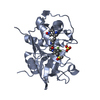
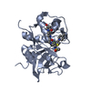
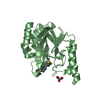
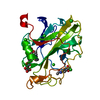
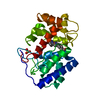
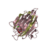
- PDB-8pch: CRYSTAL STRUCTURE OF PORCINE CATHEPSIN H DETERMINED AT 2.1 ANGSTR... -
+ Open data

- Basic information
| Entry | Database: PDB / ID: 8pch | |||||||||
|---|---|---|---|---|---|---|---|---|---|---|
| Title | CRYSTAL STRUCTURE OF PORCINE CATHEPSIN H DETERMINED AT 2.1 ANGSTROM RESOLUTION: LOCATION OF THE MINI-CHAIN C-TERMINAL CARBOXYL GROUP DEFINES CATHEPSIN H AMINOPEPTIDASE FUNCTION | |||||||||
 Components Components | (CATHEPSIN H) x 2 | |||||||||
 Keywords Keywords | HYDROLASE / PROTEASE / CYSTEINE PROTEINASE / AMINOPEPTIDASE | |||||||||
| Function / homology |  Function and homology information Function and homology informationcathepsin H / neuropeptide catabolic process / HLA-A specific activating MHC class I receptor activity / dichotomous subdivision of terminal units involved in lung branching / positive regulation of peptidase activity / Surfactant metabolism / alveolar lamellar body / immune response-regulating signaling pathway / membrane protein proteolysis / bradykinin catabolic process ...cathepsin H / neuropeptide catabolic process / HLA-A specific activating MHC class I receptor activity / dichotomous subdivision of terminal units involved in lung branching / positive regulation of peptidase activity / Surfactant metabolism / alveolar lamellar body / immune response-regulating signaling pathway / membrane protein proteolysis / bradykinin catabolic process / Neutrophil degranulation / metanephros development / surfactant homeostasis / zymogen activation / MHC class II antigen presentation / positive regulation of epithelial cell migration / thyroid hormone binding / cysteine-type endopeptidase activator activity involved in apoptotic process / aminopeptidase activity / response to retinoic acid / cysteine-type peptidase activity / ERK1 and ERK2 cascade / positive regulation of apoptotic signaling pathway / proteolysis involved in protein catabolic process / protein destabilization / T cell mediated cytotoxicity / positive regulation of angiogenesis / endopeptidase activity / lysosome / positive regulation of cell migration / immune response / serine-type endopeptidase activity / cysteine-type endopeptidase activity / positive regulation of cell population proliferation / positive regulation of gene expression / negative regulation of apoptotic process / proteolysis / extracellular space / cytosol Similarity search - Function | |||||||||
| Biological species |  | |||||||||
| Method |  X-RAY DIFFRACTION / MOLECULAR REPLACEMENT / Resolution: 2.1 Å X-RAY DIFFRACTION / MOLECULAR REPLACEMENT / Resolution: 2.1 Å | |||||||||
 Authors Authors | Guncar, G. / Podobnik, M. / Pungercar, J. / Strukelj, B. / Turk, V. / Turk, D. | |||||||||
 Citation Citation | Journal: Structure / Year: 1998 Title: Crystal structure of porcine cathepsin H determined at 2.1 A resolution: location of the mini-chain C-terminal carboxyl group defines cathepsin H aminopeptidase function. Authors: Guncar, G. / Podobnik, M. / Pungercar, J. / Strukelj, B. / Turk, V. / Turk, D. | |||||||||
| History |
|
- Structure visualization
| Structure viewer | Molecule: MolmilJmol/JSmol |
|---|
- Downloads & links
-Download
| PDBx/mmCIF format | 8pch.cif.gz | 71.8 KB | Display | PDBx/mmCIF format |
|---|---|---|---|---|
| PDB format | pdb8pch.ent.gz | 56.7 KB | Display | PDB format |
| PDBx/mmJSON format | 8pch.json.gz | Tree view | PDBx/mmJSON format | |
| Others |  Other downloads Other downloads |
-Validation report
| Summary document | 8pch_validation.pdf.gz | 433.9 KB | Display | wwPDB validaton report |
|---|---|---|---|---|
| Full document | 8pch_full_validation.pdf.gz | 434.5 KB | Display | |
| Data in XML | 8pch_validation.xml.gz | 6.4 KB | Display | |
| Data in CIF | 8pch_validation.cif.gz | 10.2 KB | Display | |
| Arichive directory | https://data.pdbj.org/pub/pdb/validation_reports/pc/8pchftp://data.pdbj.org/pub/pdb/validation_reports/pc/8pch | HTTPS FTP |
-Related structure data
| Related structure data | 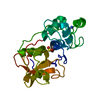 2actS S: Starting model for refinement |
|---|---|
| Similar structure data |


-Links
 PDBj
PDBj
- Assembly
| Deposited unit | 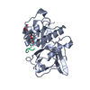
| ||||||||
|---|---|---|---|---|---|---|---|---|---|
| 1 |
| ||||||||
| Unit cell |
|
-Components
| #1: Protein | Mass: 24328.521 Da / Num. of mol.: 1 / Source method: isolated from a natural source / Source: (natural) |
|---|---|
| #2: Protein/peptide | Mass: 848.878 Da / Num. of mol.: 1 / Source method: isolated from a natural source / Source: (natural) |
| #3: Polysaccharide | beta-D-mannopyranose-(1-4)-2-acetamido-2-deoxy-beta-D-glucopyranose-(1-4)-2-acetamido-2-deoxy-beta- ...beta-D-mannopyranose-(1-4)-2-acetamido-2-deoxy-beta-D-glucopyranose-(1-4)-2-acetamido-2-deoxy-beta-D-glucopyranose Source method: isolated from a genetically manipulated source |
| #4: Water | ChemComp-HOH /  Mass: 18.015 Da / Num. of mol.: 179 / Source method: isolated from a natural source / Formula: H2O Mass: 18.015 Da / Num. of mol.: 179 / Source method: isolated from a natural source / Formula: H2O |
-Experimental details
-Experiment
| Experiment | Method: X-RAY DIFFRACTION / Number of used crystals: 1 |
|---|
- Sample preparation
| Crystal | Density Matthews: 2.27 Å3/Da / Density % sol: 45.93 % | ||||||||||||||||||||||||||||||
|---|---|---|---|---|---|---|---|---|---|---|---|---|---|---|---|---|---|---|---|---|---|---|---|---|---|---|---|---|---|---|---|
| Crystal grow | Method: vapor diffusion, sitting drop / pH: 4.8 Details: PROTEIN WAS CRYSTALLIZED USING SITTING DROP VAPOR DIFFUSION METHOD FROM 0.05M NA-ACETATE AND 5% MME PEG 5K BUFFER, PH 4.8. CONCENTRATION OF APPLIED PROTEIN WAS 11 MG/ML., vapor diffusion - sitting drop | ||||||||||||||||||||||||||||||
| Crystal grow | *PLUS pH: 5 / Method: vapor diffusion, sitting drop | ||||||||||||||||||||||||||||||
| Components of the solutions | *PLUS
|
-Data collection
| Diffraction | Mean temperature: 289 K |
|---|---|
| Diffraction source | Source: ROTATING ANODE / Type: RIGAKU RUH2R / Wavelength: 1.5418 |
| Detector | Type: MARRESEARCH / Detector: IMAGE PLATE / Date: Jul 1, 1996 / Details: MIRRORS |
| Radiation | Monochromator: MIRRORS / Monochromatic (M) / Laue (L): M / Scattering type: x-ray |
| Radiation wavelength | Wavelength: 1.5418 Å / Relative weight: 1 |
| Reflection | Highest resolution: 1.98 Å / Num. obs: 15946 / % possible obs: 95 % / Observed criterion σ(I): 1 / Rsym value: 0.118 |
| Reflection | *PLUS Highest resolution: 2.1 Å / Lowest resolution: 99 Å / % possible obs: 98 % / Num. measured all: 102384 / Rmerge(I) obs: 0.11 |
- Processing
| Software |
| ||||||||||||
|---|---|---|---|---|---|---|---|---|---|---|---|---|---|
| Refinement | Method to determine structure: MOLECULAR REPLACEMENT Starting model: 2ACT Resolution: 2.1→8 Å / Cross valid method: R-FREE,KICKED OMIT MAP / σ(F): 1
| ||||||||||||
| Refinement step | Cycle: LAST / Resolution: 2.1→8 Å
| ||||||||||||
| Refinement | *PLUS Rfactor obs: 0.198 | ||||||||||||
| Solvent computation | *PLUS | ||||||||||||
| Displacement parameters | *PLUS Biso mean: 13.7 Å2 | ||||||||||||
| Refine LS restraints | *PLUS
|
