 Movie
Movie Controller

[English] 日本語
 Yorodumi
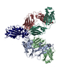
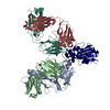
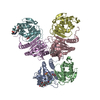
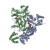
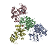
- PDB-7che: Crystal structure of the SARS-CoV-2 RBD in complex with BD-236 Fa... -
+ Open data

- Basic information
| Entry | Database: PDB / ID: 7che | ||||||
|---|---|---|---|---|---|---|---|
| Title | Crystal structure of the SARS-CoV-2 RBD in complex with BD-236 Fab and BD-368-2 Fab | ||||||
 Components Components |
| ||||||
 Keywords Keywords |  ANTIVIRAL PROTEIN/IMMUNE SYSTEM / mAb / ANTIVIRAL PROTEIN-IMMUNE SYSTEM complex ANTIVIRAL PROTEIN/IMMUNE SYSTEM / mAb / ANTIVIRAL PROTEIN-IMMUNE SYSTEM complex | ||||||
| Function / homology |  Function and homology information Function and homology informationMaturation of spike protein / viral translation / Translation of Structural Proteins / Virion Assembly and Release / host cell surface / host extracellular space / suppression by virus of host tetherin activity / Induction of Cell-Cell Fusion / structural constituent of virion / host cell endoplasmic reticulum-Golgi intermediate compartment membrane ...Maturation of spike protein / viral translation / Translation of Structural Proteins / Virion Assembly and Release / host cell surface / host extracellular space / suppression by virus of host tetherin activity / Induction of Cell-Cell Fusion / structural constituent of virion / host cell endoplasmic reticulum-Golgi intermediate compartment membrane / entry receptor-mediated virion attachment to host cell / receptor-mediated endocytosis of virus by host cell / Attachment and Entry / membrane fusion / positive regulation of viral entry into host cell / receptor-mediated virion attachment to host cell / receptor ligand activity / host cell surface receptor binding / fusion of virus membrane with host plasma membrane / fusion of virus membrane with host endosome membrane / viral envelope / symbiont-mediated suppression of host type I interferon-mediated signaling pathway / virion attachment to host cell / SARS-CoV-2 activates/modulates innate and adaptive immune responses / host cell plasma membrane / virion membrane / membrane / identical protein binding / plasma membraneSimilarity search - Function | ||||||
| Biological species |  Homo sapiens (human) Homo sapiens (human)  Severe acute respiratory syndrome coronavirus 2 Severe acute respiratory syndrome coronavirus 2 | ||||||
| Method | X-RAY DIFFRACTION / SYNCHROTRON / MOLECULAR REPLACEMENT / Resolution: 3.416 Å | ||||||
 Authors Authors | Xiao, J. / Zhu, Q. | ||||||
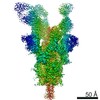
 Citation Citation | Journal: Cell / Year: 2020 Title: Structurally Resolved SARS-CoV-2 Antibody Shows High Efficacy in Severely Infected Hamsters and Provides a Potent Cocktail Pairing Strategy. Authors: Shuo Du / Yunlong Cao / Qinyu Zhu / Pin Yu / Feifei Qi / Guopeng Wang / Xiaoxia Du / Linlin Bao / Wei Deng / Hua Zhu / Jiangning Liu / Jianhui Nie / Yinghui Zheng / Haoyu Liang / Ruixue Liu ...Authors: Shuo Du / Yunlong Cao / Qinyu Zhu / Pin Yu / Feifei Qi / Guopeng Wang / Xiaoxia Du / Linlin Bao / Wei Deng / Hua Zhu / Jiangning Liu / Jianhui Nie / Yinghui Zheng / Haoyu Liang / Ruixue Liu / Shuran Gong / Hua Xu / Ayijiang Yisimayi / Qi Lv / Bo Wang / Runsheng He / Yunlin Han / Wenjie Zhao / Yali Bai / Yajin Qu / Xiang Gao / Chenggong Ji / Qisheng Wang / Ning Gao / Weijin Huang / Youchun Wang / X Sunney Xie / Xiao-Dong Su / Junyu Xiao / Chuan Qin /  Abstract: Understanding how potent neutralizing antibodies (NAbs) inhibit SARS-CoV-2 is critical for effective therapeutic development. We previously described BD-368-2, a SARS-CoV-2 NAb with high potency; ...Understanding how potent neutralizing antibodies (NAbs) inhibit SARS-CoV-2 is critical for effective therapeutic development. We previously described BD-368-2, a SARS-CoV-2 NAb with high potency; however, its neutralization mechanism is largely unknown. Here, we report the 3.5-Å cryo-EM structure of BD-368-2/trimeric-spike complex, revealing that BD-368-2 fully blocks ACE2 recognition by occupying all three receptor-binding domains (RBDs) simultaneously, regardless of their "up" or "down" conformations. Also, BD-368-2 treats infected adult hamsters at low dosages and at various administering windows, in contrast to placebo hamsters that manifested severe interstitial pneumonia. Moreover, BD-368-2's epitope completely avoids the common binding site of VH3-53/VH3-66 recurrent NAbs, evidenced by tripartite co-crystal structures with RBDs. Pairing BD-368-2 with a potent recurrent NAb neutralizes SARS-CoV-2 pseudovirus at pM level and rescues mutation-induced neutralization escapes. Together, our results rationalized a new RBD epitope that leads to high neutralization potency and demonstrated BD-368-2's therapeutic potential in treating COVID-19. | ||||||
| History |
|
- Structure visualization
| Structure viewer | Molecule: MolmilJmol/JSmol |
|---|
- Downloads & links
-Download
| PDBx/mmCIF format | 7che.cif.gz | 214.4 KB | Display | PDBx/mmCIF format |
|---|---|---|---|---|
| PDB format | pdb7che.ent.gz | 168 KB | Display | PDB format |
| PDBx/mmJSON format | 7che.json.gz | Tree view | PDBx/mmJSON format | |
| Others |  Other downloads Other downloads |
-Validation report
| Arichive directory | https://data.pdbj.org/pub/pdb/validation_reports/ch/7cheftp://data.pdbj.org/pub/pdb/validation_reports/ch/7che | HTTPS FTP |
|---|
-Related structure data
| Related structure data |  7ch4C 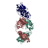 7ch5C  7chbC  7chcC  7chfC 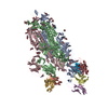 7chhC 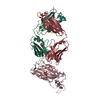 6w41S S: Starting model for refinement C: citing same article ( |
|---|---|
| Similar structure data |


-Links
 PDBj
PDBj


- Assembly
| Deposited unit | 
| ||||||||
|---|---|---|---|---|---|---|---|---|---|
| 1 |
| ||||||||
| Unit cell |
|
-Components
| #1: Antibody | Mass: 23333.113 Da / Num. of mol.: 1 Source method: isolated from a genetically manipulated source Source: (gene. exp.) Homo sapiens (human) / Cell line (production host): 293F / Production host: Homo sapiens (human) |
|---|---|
| #2: Antibody | Mass: 23100.668 Da / Num. of mol.: 1 Source method: isolated from a genetically manipulated source Source: (gene. exp.) Homo sapiens (human) / Cell line (production host): 293F / Production host: Homo sapiens (human) |
| #3: Protein | Mass: 25122.336 Da / Num. of mol.: 1 Source method: isolated from a genetically manipulated source Source: (gene. exp.) Severe acute respiratory syndrome coronavirus 2Gene: S, 2 / Production host:  Trichoplusia ni (cabbage looper) / References: UniProt: P0DTC2 Trichoplusia ni (cabbage looper) / References: UniProt: P0DTC2 |
| #4: Antibody | Mass: 24355.225 Da / Num. of mol.: 1 Source method: isolated from a genetically manipulated source Source: (gene. exp.) Homo sapiens (human) / Cell line (production host): 293F / Production host: Homo sapiens (human) |
| #5: Antibody | Mass: 23902.590 Da / Num. of mol.: 1 Source method: isolated from a genetically manipulated source Source: (gene. exp.) Homo sapiens (human) / Cell line (production host): 293F / Production host: Homo sapiens (human) |
-Experimental details
-Experiment
| Experiment | Method: X-RAY DIFFRACTION / Number of used crystals: 1 |
|---|
- Sample preparation
| Crystal | Density Matthews: 2.79 Å3/Da / Density % sol: 55.91 % |
|---|---|
| Crystal grow | Temperature: 293 K / Method: vapor diffusion, sitting drop Details: 0.1M sodium acetate pH 4.0 and 10% (w/v) Polyethylene glycol monomethyl ether 2,000 |
-Data collection
| Diffraction | Mean temperature: 80 K / Serial crystal experiment: N |
|---|---|
| Diffraction source | Source: SYNCHROTRON / Site: SSRF / Beamline: BL17U1 / Wavelength: 0.97918 Å |
| Detector | Type: DECTRIS EIGER X 16M / Detector: PIXEL / Date: Jun 25, 2020 |
| Radiation | Protocol: SINGLE WAVELENGTH / Monochromatic (M) / Laue (L): M / Scattering type: x-ray |
| Radiation wavelength | Wavelength: 0.97918 Å / Relative weight: 1 |
| Reflection | Resolution: 3.416→50 Å / Num. obs: 17742 / % possible obs: 94.54 % / Redundancy: 12.7 % / Biso Wilson estimate: 64.08 Å2 / CC1/2: 0.98 / Net I/σ(I): 11.55 |
| Reflection shell | Resolution: 3.45→3.51 Å / Num. unique obs: 1036 / CC1/2: 0.692 |
- Processing
| Software |
| ||||||||||||||||||||||||||||||||||||||||||||||||||||||||||||||||||||||||||||||||||||
|---|---|---|---|---|---|---|---|---|---|---|---|---|---|---|---|---|---|---|---|---|---|---|---|---|---|---|---|---|---|---|---|---|---|---|---|---|---|---|---|---|---|---|---|---|---|---|---|---|---|---|---|---|---|---|---|---|---|---|---|---|---|---|---|---|---|---|---|---|---|---|---|---|---|---|---|---|---|---|---|---|---|---|---|---|---|
| Refinement | Method to determine structure: MOLECULAR REPLACEMENT Starting model: 6W41 Resolution: 3.416→49.619 Å / SU ML: 0.4 / Cross valid method: THROUGHOUT / σ(F): 1.35 / Phase error: 23.55 / Stereochemistry target values: ML
| ||||||||||||||||||||||||||||||||||||||||||||||||||||||||||||||||||||||||||||||||||||
| Solvent computation | Shrinkage radii: 0.9 Å / VDW probe radii: 1.11 Å / Solvent model: FLAT BULK SOLVENT MODEL | ||||||||||||||||||||||||||||||||||||||||||||||||||||||||||||||||||||||||||||||||||||
| Displacement parameters | Biso max: 145.61 Å2 / Biso mean: 54.7801 Å2 / Biso min: 15.58 Å2 | ||||||||||||||||||||||||||||||||||||||||||||||||||||||||||||||||||||||||||||||||||||
| Refinement step | Cycle: final / Resolution: 3.416→49.619 Å
| ||||||||||||||||||||||||||||||||||||||||||||||||||||||||||||||||||||||||||||||||||||
| Refine LS restraints |
| ||||||||||||||||||||||||||||||||||||||||||||||||||||||||||||||||||||||||||||||||||||
| LS refinement shell | Refine-ID: X-RAY DIFFRACTION / Rfactor Rfree error: 0
|
