 Movie
Movie Controller

[English] 日本語
 Yorodumi
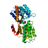
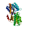
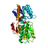
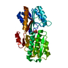
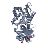
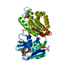
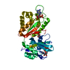
- PDB-7abp: SUGAR-BINDING AND CRYSTALLOGRAPHIC STUDIES OF AN ARABINOSE-BINDIN... -
+ Open data

- Basic information
| Entry | Database: PDB / ID: 7abp | |||||||||
|---|---|---|---|---|---|---|---|---|---|---|
| Title | SUGAR-BINDING AND CRYSTALLOGRAPHIC STUDIES OF AN ARABINOSE-BINDING PROTEIN MUTANT (MET108LEU) WHICH EXHIBITS ENHANCED AFFINITY AND ALTERED SPECIFICITY | |||||||||
 Components Components | L-ARABINOSE-BINDING PROTEIN | |||||||||
 Keywords Keywords | BINDING PROTEINS | |||||||||
| Function / homology |  Function and homology information Function and homology informationABC-type monosaccharide transporter activity / L-arabinose transmembrane transport / monosaccharide binding / ATP-binding cassette (ABC) transporter complex, substrate-binding subunit-containing / outer membrane-bounded periplasmic space / carbohydrate binding / membrane Similarity search - Function | |||||||||
| Biological species |  | |||||||||
| Method |  X-RAY DIFFRACTION / Resolution: 1.67 Å X-RAY DIFFRACTION / Resolution: 1.67 Å | |||||||||
 Authors Authors | Vermersch, P.S. / Tesmer, J.J.G. / Quiocho, F.A. | |||||||||
 Citation Citation | Journal: Biochemistry / Year: 1991 Title: Sugar-binding and crystallographic studies of an arabinose-binding protein mutant (Met108Leu) that exhibits enhanced affinity and altered specificity. Authors: Vermersch, P.S. / Lemon, D.D. / Tesmer, J.J. / Quiocho, F.A. #1: Journal: Nature / Year: 1989Title: Substrate Specificity and Affinity of a Protein Modulated by Bound Water Molecules Authors: Quiocho, F.A. / Wilson, D.K. / Vyas, N.K. #2: Journal: Nature / Year: 1984Title: Novel Stereospecificity of the L-Arabinose-Binding Protein Authors: Quiocho, F.A. / Vyas, N.K. #3: Journal: J.Biol.Chem. / Year: 1982Title: Hinge-Bending in L-Arabinose-Binding Protein. The "Venus'S-Flytrap" Model Authors: Mao, B. / Pear, M.R. / Mccammon, J.A. / Quiocho, F.A. #4: Journal: J.Mol.Biol. / Year: 1981Title: Structure of the L-Arabinose-Binding Protein from Escherichia Coli at 2.4 Angstroms Resolution Authors: Gilliland, G.L. / Quiocho, F.A. #5: Journal: J.Biol.Chem. / Year: 1981Title: L-Arabinose-Binding Protein-Sugar Complex at 2.4 Angstroms Resolution. Stereochemistry and Evidence for a Structural Change Authors: Newcomer, M.E. / Gilliand, G.L. / Quiocho, F.A. #6: Journal: J.Biol.Chem. / Year: 1981Title: The Radius of Gyration of L-Arabinose-Binding Protein Decreases Upon Binding of Ligand Authors: Newcomer, M.E. / Lewis, B.A. / Quiocho, F.A. #7: Journal: J.Biol.Chem. / Year: 1979Title: The Thiol Group of the L-Arabinose-Binding Protein. Chromophoric Labeling and Chemical Identification of the Sugar-Binding Site Authors: Miller /III, D.M. / Newcomer, M.E. / Quiocho, F.A. #8: Journal: J.Biol.Chem. / Year: 1979Title: Location of the Sugar-Binding Site of L-Arabinose-Binding Protein. Sugar Derivative Syntheses, Sugar Binding Specificity, and Difference Fourier Analyses Authors: Newcomer, M.E. / Miller /III, D.M. / Quiocho, F.A. #9: Journal: J.Biol.Chem. / Year: 1977Title: The 2.8-Angstroms Resolution Structure of the L-Arabinose-Binding Protein from Escherichia Coli Authors: Quiocho, F.A. / Gilliland, G.L. / Phillips Jr., G.N. #10: Journal: Proc.Natl.Acad.Sci.USA / Year: 1976Title: Structure of L-Arabinose-Binding Protein from Escherichia Coli at 5 Angstroms Resolution and Preliminary Results at 3.5 Angstroms Authors: Phillips Jr., G.N. / Mahajan, V.K. / Siu, A.K.Q. / Quiocho, F.A. | |||||||||
| History |
|
- Structure visualization
| Structure viewer | Molecule: MolmilJmol/JSmol |
|---|
- Downloads & links
-Download
| PDBx/mmCIF format | 7abp.cif.gz | 74.7 KB | Display | PDBx/mmCIF format |
|---|---|---|---|---|
| PDB format | pdb7abp.ent.gz | 55.4 KB | Display | PDB format |
| PDBx/mmJSON format | 7abp.json.gz | Tree view | PDBx/mmJSON format | |
| Others |  Other downloads Other downloads |
-Validation report
| Arichive directory | https://data.pdbj.org/pub/pdb/validation_reports/ab/7abpftp://data.pdbj.org/pub/pdb/validation_reports/ab/7abp | HTTPS FTP |
|---|
-Related structure data


-Links
 PDBj
PDBj- Assembly
| Deposited unit | 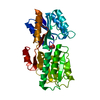
| ||||||||
|---|---|---|---|---|---|---|---|---|---|
| 1 |
| ||||||||
| Unit cell |
|
-Components
| #1: Protein | Mass: 33232.957 Da / Num. of mol.: 1 Source method: isolated from a genetically manipulated source Source: (gene. exp.) |
|---|---|
| #2: Sugar | ChemComp-FCA /   Type: D-saccharide, alpha linking / Mass: 164.156 Da / Num. of mol.: 1 Type: D-saccharide, alpha linking / Mass: 164.156 Da / Num. of mol.: 1Source method: isolated from a genetically manipulated source Formula: C6H12O5 |
| #3: Sugar | ChemComp-FCB /   Type: D-saccharide, beta linking / Mass: 164.156 Da / Num. of mol.: 1 Type: D-saccharide, beta linking / Mass: 164.156 Da / Num. of mol.: 1Source method: isolated from a genetically manipulated source Formula: C6H12O5 |
| #4: Water | ChemComp-HOH /  Mass: 18.015 Da / Num. of mol.: 193 Mass: 18.015 Da / Num. of mol.: 193Source method: isolated from a genetically manipulated source Formula: H2O |
-Experimental details
-Experiment
| Experiment | Method: X-RAY DIFFRACTION |
|---|
- Sample preparation
| Crystal | Density Matthews: 2.35 Å3/Da / Density % sol: 47.72 % | ||||||||||||||||||||||||||||||||||||||||||||||||||||||||
|---|---|---|---|---|---|---|---|---|---|---|---|---|---|---|---|---|---|---|---|---|---|---|---|---|---|---|---|---|---|---|---|---|---|---|---|---|---|---|---|---|---|---|---|---|---|---|---|---|---|---|---|---|---|---|---|---|---|
| Crystal grow | *PLUS Temperature: 4 ℃ / pH: 5 / Method: vapor diffusion, hanging drop / Details: referred to J.Biol.Chem. 265.16592-16603 | ||||||||||||||||||||||||||||||||||||||||||||||||||||||||
| Components of the solutions | *PLUS
|
-Data collection
| Reflection | *PLUS Highest resolution: 1.67 Å / Lowest resolution: 8 Å / Num. obs: 33726 / % possible obs: 91 % / Num. measured all: 120410 / Rmerge(I) obs: 0.0595 |
|---|
- Processing
| Software | Name: PROLSQ / Classification: refinement | |||||||||||||||||||||||||||||||||||||||||||||||||||||||||||||||
|---|---|---|---|---|---|---|---|---|---|---|---|---|---|---|---|---|---|---|---|---|---|---|---|---|---|---|---|---|---|---|---|---|---|---|---|---|---|---|---|---|---|---|---|---|---|---|---|---|---|---|---|---|---|---|---|---|---|---|---|---|---|---|---|---|
| Refinement | Resolution: 1.67→8 Å /
| |||||||||||||||||||||||||||||||||||||||||||||||||||||||||||||||
| Refinement step | Cycle: LAST / Resolution: 1.67→8 Å
| |||||||||||||||||||||||||||||||||||||||||||||||||||||||||||||||
| Refine LS restraints |
| |||||||||||||||||||||||||||||||||||||||||||||||||||||||||||||||
| Refinement | *PLUS Highest resolution: 1.67 Å / Lowest resolution: 8 Å / σ(F): 2 / Rfactor obs: 0.162 / Num. reflection obs: 29509 | |||||||||||||||||||||||||||||||||||||||||||||||||||||||||||||||
| Solvent computation | *PLUS | |||||||||||||||||||||||||||||||||||||||||||||||||||||||||||||||
| Displacement parameters | *PLUS | |||||||||||||||||||||||||||||||||||||||||||||||||||||||||||||||
| Refine LS restraints | *PLUS
|
