 Movie
Movie Controller

[English] 日本語
 Yorodumi
- PDB-6t92: NAD+-dependent fungal formate dehydrogenase from Chaetomium therm... -
+ Open data

- Basic information
| Entry | Database: PDB / ID: 6t92 | ||||||
|---|---|---|---|---|---|---|---|
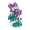
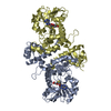
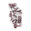
| Title | NAD+-dependent fungal formate dehydrogenase from Chaetomium thermophilum: A complex of N120C mutant protein with the reduced form of the cofactor NADH and the substrate formate at a secondary site. | ||||||
 Components Components | Formate dehydrogenase | ||||||
 Keywords Keywords | CYTOSOLIC PROTEIN / NAD+ | ||||||
| Function / homology |  Function and homology information Function and homology informationformate catabolic process / formate dehydrogenase / formate dehydrogenase (NAD+) activity / oxidoreductase activity, acting on the CH-OH group of donors, NAD or NADP as acceptor / NAD binding / cytosol Similarity search - Function | ||||||
| Biological species |  Chaetomium thermophilum (fungus) Chaetomium thermophilum (fungus) | ||||||
| Method |  X-RAY DIFFRACTION / SYNCHROTRON / MOLECULAR REPLACEMENT / Resolution: 1.12 Å X-RAY DIFFRACTION / SYNCHROTRON / MOLECULAR REPLACEMENT / Resolution: 1.12 Å | ||||||
 Authors Authors | Isupov, M.N. / Yelmazer, B. / De Rose, S.A. / Littlechild, J.A. | ||||||
 Citation Citation | Journal: J.Struct.Biol. / Year: 2020 Title: Structural insights into the NAD + -dependent formate dehydrogenase mechanism revealed from the NADH complex and the formate NAD + ternary complex of the Chaetomium thermophilum enzyme. Authors: Yilmazer, B. / Isupov, M.N. / De Rose, S.A. / Bulut, H. / Benninghoff, J.C. / Binay, B. / Littlechild, J.A. | ||||||
| History |
|
- Structure visualization
| Structure viewer | Molecule: MolmilJmol/JSmol |
|---|
- Downloads & links
-Download
| PDBx/mmCIF format | 6t92.cif.gz | 616.6 KB | Display | PDBx/mmCIF format |
|---|---|---|---|---|
| PDB format | pdb6t92.ent.gz | Display | PDB format | |
| PDBx/mmJSON format | 6t92.json.gz | Tree view | PDBx/mmJSON format | |
| Others |  Other downloads Other downloads |
-Validation report
| Arichive directory | https://data.pdbj.org/pub/pdb/validation_reports/t9/6t92ftp://data.pdbj.org/pub/pdb/validation_reports/t9/6t92 | HTTPS FTP |
|---|
-Related structure data
| Related structure data |  6t8yC  6t8zC  6t94C  5dnaS S: Starting model for refinement C: citing same article ( |
|---|---|
| Similar structure data |

-Links
 PDBj
PDBj

- Assembly
| Deposited unit | 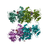
| ||||||||||||||||||||||||||||
|---|---|---|---|---|---|---|---|---|---|---|---|---|---|---|---|---|---|---|---|---|---|---|---|---|---|---|---|---|---|
| 1 | 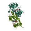
| ||||||||||||||||||||||||||||
| 2 | 
| ||||||||||||||||||||||||||||
| Unit cell |
| ||||||||||||||||||||||||||||
| Noncrystallographic symmetry (NCS) | NCS domain:
NCS ensembles :
|
-Components
-Protein , 1 types, 4 molecules AAABBBCCCDDD
| #1: Protein | Mass: 45630.441 Da / Num. of mol.: 4 / Mutation: N120C Source method: isolated from a genetically manipulated source Source: (gene. exp.) Chaetomium thermophilum (strain DSM 1495 / CBS 144.50 / IMI 039719) (fungus)Strain: DSM 1495 / CBS 144.50 / IMI 039719 / Gene: FDH, CTHT_0067590 / Production host:  |
|---|
-Non-polymers , 5 types, 1781 molecules 


| #2: Chemical | ChemComp-PEG /  Mass: 106.120 Da / Num. of mol.: 5 / Source method: obtained synthetically / Formula: C4H10O3 Mass: 106.120 Da / Num. of mol.: 5 / Source method: obtained synthetically / Formula: C4H10O3#3: Chemical | ChemComp-NAI /  Mass: 665.441 Da / Num. of mol.: 4 / Source method: obtained synthetically / Formula: C21H29N7O14P2 / Feature type: SUBJECT OF INVESTIGATION Mass: 665.441 Da / Num. of mol.: 4 / Source method: obtained synthetically / Formula: C21H29N7O14P2 / Feature type: SUBJECT OF INVESTIGATION#4: Chemical | ChemComp-FMT /  Mass: 46.025 Da / Num. of mol.: 7 / Source method: obtained synthetically / Formula: CH2O2 / Feature type: SUBJECT OF INVESTIGATION Mass: 46.025 Da / Num. of mol.: 7 / Source method: obtained synthetically / Formula: CH2O2 / Feature type: SUBJECT OF INVESTIGATION#5: Chemical | ChemComp-EDO /  Mass: 62.068 Da / Num. of mol.: 6 / Source method: obtained synthetically / Formula: C2H6O2 Mass: 62.068 Da / Num. of mol.: 6 / Source method: obtained synthetically / Formula: C2H6O2#6: Water | ChemComp-HOH / | Mass: 18.015 Da / Num. of mol.: 1759 / Source method: isolated from a natural source / Formula: H2O |
|---|
-Details
| Has ligand of interest | Y |
|---|
-Experimental details
-Experiment
| Experiment | Method: X-RAY DIFFRACTION / Number of used crystals: 1 |
|---|
- Sample preparation
| Crystal | Density Matthews: 2.45 Å3/Da / Density % sol: 49.88 % |
|---|---|
| Crystal grow | Temperature: 291 K / Method: microbatch / pH: 7.5 Details: 0.2 M Magnesium chloride hexahydrate, 0.1 M Tris pH 7.5, 10 % w/v PEG 1000, 10 % w/v PEG 8000 |
-Data collection
| Diffraction | Mean temperature: 100 K / Serial crystal experiment: N |
|---|---|
| Diffraction source | Source: SYNCHROTRON / Site: Diamond  / Beamline: I03 / Wavelength: 0.9763 Å / Beamline: I03 / Wavelength: 0.9763 Å |
| Detector | Type: DECTRIS PILATUS3 6M / Detector: PIXEL / Date: Feb 26, 2018 |
| Radiation | Protocol: SINGLE WAVELENGTH / Monochromatic (M) / Laue (L): M / Scattering type: x-ray |
| Radiation wavelength | Wavelength: 0.9763 Å / Relative weight: 1 |
| Reflection | Resolution: 1.12→63.93 Å / Num. obs: 548213 / % possible obs: 82.1 % / Redundancy: 1.7 % / Biso Wilson estimate: 15.7 Å2 / CC1/2: 0.998 / Net I/σ(I): 5.7 |
| Reflection shell | Resolution: 1.12→1.14 Å / Redundancy: 1.4 % / Mean I/σ(I) obs: 0.8 / Num. unique obs: 17604 / CC1/2: 0.291 / % possible all: 53.2 |
- Processing
| Software |
| ||||||||||||||||||||||||||||||||||||||||||||||||||||||||||||||||||||||||||||||||||||||||||||||||||||||||||||||||||||||||||||||||||||||||||||||||||||||||||||||||||||||||||||||||||||||
|---|---|---|---|---|---|---|---|---|---|---|---|---|---|---|---|---|---|---|---|---|---|---|---|---|---|---|---|---|---|---|---|---|---|---|---|---|---|---|---|---|---|---|---|---|---|---|---|---|---|---|---|---|---|---|---|---|---|---|---|---|---|---|---|---|---|---|---|---|---|---|---|---|---|---|---|---|---|---|---|---|---|---|---|---|---|---|---|---|---|---|---|---|---|---|---|---|---|---|---|---|---|---|---|---|---|---|---|---|---|---|---|---|---|---|---|---|---|---|---|---|---|---|---|---|---|---|---|---|---|---|---|---|---|---|---|---|---|---|---|---|---|---|---|---|---|---|---|---|---|---|---|---|---|---|---|---|---|---|---|---|---|---|---|---|---|---|---|---|---|---|---|---|---|---|---|---|---|---|---|---|---|---|---|
| Refinement | Method to determine structure: MOLECULAR REPLACEMENT Starting model: 5dna Resolution: 1.12→63.93 Å / Cor.coef. Fo:Fc: 0.966 / Cor.coef. Fo:Fc free: 0.958 / Cross valid method: FREE R-VALUE / ESU R: 0.044 / ESU R Free: 0.046 Details: Hydrogens have been added in their riding positions
| ||||||||||||||||||||||||||||||||||||||||||||||||||||||||||||||||||||||||||||||||||||||||||||||||||||||||||||||||||||||||||||||||||||||||||||||||||||||||||||||||||||||||||||||||||||||
| Solvent computation | Ion probe radii: 0.8 Å / Shrinkage radii: 0.8 Å / VDW probe radii: 1.2 Å | ||||||||||||||||||||||||||||||||||||||||||||||||||||||||||||||||||||||||||||||||||||||||||||||||||||||||||||||||||||||||||||||||||||||||||||||||||||||||||||||||||||||||||||||||||||||
| Displacement parameters | Biso mean: 18.109 Å2
| ||||||||||||||||||||||||||||||||||||||||||||||||||||||||||||||||||||||||||||||||||||||||||||||||||||||||||||||||||||||||||||||||||||||||||||||||||||||||||||||||||||||||||||||||||||||
| Refinement step | Cycle: LAST / Resolution: 1.12→63.93 Å
| ||||||||||||||||||||||||||||||||||||||||||||||||||||||||||||||||||||||||||||||||||||||||||||||||||||||||||||||||||||||||||||||||||||||||||||||||||||||||||||||||||||||||||||||||||||||
| Refine LS restraints |
|