 Movie
Movie Controller


+ Open data

- Basic information
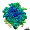
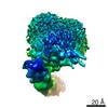
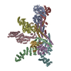
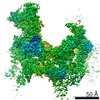
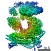
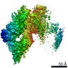
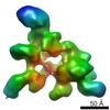
| Entry | Database: PDB / ID: 6sxo | ||||||
|---|---|---|---|---|---|---|---|
| Title | Cryo-EM structure of the human Ebp1-ribosome complex | ||||||
 Components Components |
| ||||||
 Keywords Keywords |  RIBOSOME / Human Ebp1 / PA2G4 family / MET-AP homolog / expansion segment ES27L RIBOSOME / Human Ebp1 / PA2G4 family / MET-AP homolog / expansion segment ES27L | ||||||
| Function / homology |  Function and homology information Function and homology informationeukaryotic 80S initiation complex / axial mesoderm development / positive regulation of intrinsic apoptotic signaling pathway in response to DNA damage by p53 class mediator / regulation of translation involved in cellular response to UV / 90S preribosome assembly / positive regulation of DNA damage response, signal transduction by p53 class mediator resulting in transcription of p21 class mediator / TORC2 complex binding / middle ear morphogenesis / Peptide chain elongation / Selenocysteine synthesis ...eukaryotic 80S initiation complex / axial mesoderm development / positive regulation of intrinsic apoptotic signaling pathway in response to DNA damage by p53 class mediator / regulation of translation involved in cellular response to UV / 90S preribosome assembly / positive regulation of DNA damage response, signal transduction by p53 class mediator resulting in transcription of p21 class mediator / TORC2 complex binding / middle ear morphogenesis / Peptide chain elongation / Selenocysteine synthesis / protein-RNA complex assembly / Formation of a pool of free 40S subunits / Eukaryotic Translation Termination / Response of EIF2AK4 (GCN2) to amino acid deficiency / SRP-dependent cotranslational protein targeting to membrane / Viral mRNA Translation / Nonsense Mediated Decay (NMD) independent of the Exon Junction Complex (EJC) / GTP hydrolysis and joining of the 60S ribosomal subunit / L13a-mediated translational silencing of Ceruloplasmin expression / Major pathway of rRNA processing in the nucleolus and cytosol / Nonsense Mediated Decay (NMD) enhanced by the Exon Junction Complex (EJC) / DNA damage response, signal transduction by p53 class mediator resulting in cell cycle arrest / maturation of LSU-rRNA from tricistronic rRNA transcript (SSU-rRNA, 5.8S rRNA, LSU-rRNA) / ribosomal large subunit biogenesis / ossification / cytosolic ribosome / positive regulation of translation / skeletal system development / positive regulation of cell differentiation / sensory perception of sound / cellular response to gamma radiation / mRNA 5'-UTR binding / rRNA processing / Regulation of expression of SLITs and ROBOs / transcription corepressor activity / cellular response to UV / azurophil granule lumen / regulation of translation / cytoplasmic translation / cytosolic large ribosomal subunit / postsynaptic density / nucleic acid binding / rRNA binding / structural constituent of ribosome / cadherin binding / ribonucleoprotein complex / translation / focal adhesion / mRNA binding / negative regulation of DNA-templated transcription / synapse / ubiquitin protein ligase binding / Neutrophil degranulation / nucleolus / negative regulation of apoptotic process / RNA binding / extracellular exosome / extracellular region / nucleoplasm / membrane / nucleus / cytosol / cytoplasmSimilarity search - Function | ||||||
| Biological species |  Homo sapiens (human) Homo sapiens (human) | ||||||
| Method | ELECTRON MICROSCOPY / single particle reconstruction / cryo EM / Resolution: 3.3 Å | ||||||
 Authors Authors | Wild, K. / Aleksic, M. / Pfeffer, M. / Sinning, I. | ||||||
| Funding support |  Germany, 1items Germany, 1items
| ||||||
 Citation Citation | Journal: Nat Commun / Year: 2020 Title: MetAP-like Ebp1 occupies the human ribosomal tunnel exit and recruits flexible rRNA expansion segments. Authors: Klemens Wild / Milan Aleksić / Karine Lapouge / Keven D Juaire / Dirk Flemming / Stefan Pfeffer / Irmgard Sinning / Abstract: Human Ebp1 is a member of the proliferation-associated 2G4 (PA2G4) family and plays an important role in cancer regulation. Ebp1 shares the methionine aminopeptidase (MetAP) fold and binds to mature ...Human Ebp1 is a member of the proliferation-associated 2G4 (PA2G4) family and plays an important role in cancer regulation. Ebp1 shares the methionine aminopeptidase (MetAP) fold and binds to mature 80S ribosomes for translational control. Here, we present a cryo-EM single particle analysis reconstruction of Ebp1 bound to non-translating human 80S ribosomes at a resolution range from 3.3 to ~8 Å. Ebp1 blocks the tunnel exit with major interactions to the general uL23/uL29 docking site for nascent chain-associated factors complemented by eukaryote-specific eL19 and rRNA helix H59. H59 is defined as dynamic adaptor undergoing significant remodeling upon Ebp1 binding. Ebp1 recruits rRNA expansion segment ES27L to the tunnel exit via specific interactions with rRNA consensus sequences. The Ebp1-ribosome complex serves as a template for MetAP binding and provides insights into the structural principles for spatial coordination of co-translational events and molecular triage at the ribosomal tunnel exit. | ||||||
| History |
|
- Structure visualization
| Movie |
Movie viewer |
|---|---|
| Structure viewer | Molecule: MolmilJmol/JSmol |
- Downloads & links
-Download
| PDBx/mmCIF format | 6sxo.cif.gz | 426.8 KB | Display | PDBx/mmCIF format |
|---|---|---|---|---|
| PDB format | pdb6sxo.ent.gz | Display | PDB format | |
| PDBx/mmJSON format | 6sxo.json.gz | Tree view | PDBx/mmJSON format | |
| Others |  Other downloads Other downloads |
-Validation report
| Arichive directory | https://data.pdbj.org/pub/pdb/validation_reports/sx/6sxoftp://data.pdbj.org/pub/pdb/validation_reports/sx/6sxo | HTTPS FTP |
|---|
-Related structure data
| Related structure data |  10344MC C: citing same article ( M: map data used to model this data |
|---|---|
| Similar structure data |


-Links
 PDBj
PDBj


- Assembly
| Deposited unit | 
|
|---|---|
| 1 |
|
-Components
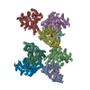
-Protein , 1 types, 1 molecules A
| #1: Protein | Mass: 43851.879 Da / Num. of mol.: 1 Source method: isolated from a genetically manipulated source Source: (gene. exp.) Homo sapiens (human) / Gene: PA2G4, EBP1 / Production host:  Escherichia coli (E. coli) / References: UniProt: Q9UQ80 Escherichia coli (E. coli) / References: UniProt: Q9UQ80 |
|---|
-60S ribosomal protein ... , 5 types, 5 molecules LhLkLRLXLY
| #2: Protein | / Large ribosomal subunit protein uL29 Mass: 14593.624 Da / Num. of mol.: 1 / Source method: isolated from a natural source / Source: (natural) Homo sapiens (human) / References: UniProt: P42766 |
|---|---|
| #3: Protein | / Large ribosomal subunit protein eL38 Mass: 8238.948 Da / Num. of mol.: 1 / Source method: isolated from a natural source / Source: (natural) Homo sapiens (human) / References: UniProt: P63173 |
| #4: Protein | / Large ribosomal subunit protein eL19 Mass: 23535.281 Da / Num. of mol.: 1 / Source method: isolated from a natural source / Source: (natural) Homo sapiens (human) / References: UniProt: P84098 |
| #5: Protein | / Large ribosomal subunit protein uL23 Mass: 17740.193 Da / Num. of mol.: 1 / Source method: isolated from a natural source / Source: (natural) Homo sapiens (human) / References: UniProt: P62750 |
| #6: Protein | / Large ribosomal subunit protein uL24 Mass: 17303.363 Da / Num. of mol.: 1 / Source method: isolated from a natural source / Source: (natural) Homo sapiens (human) / References: UniProt: P61254 |
-RNA chain , 2 types, 2 molecules L5L8
| #7: RNA chain | Mass: 1640222.125 Da / Num. of mol.: 1 / Source method: isolated from a natural source / Source: (natural) Homo sapiens (human) / References: GenBank: 86475748 |
|---|---|
| #8: RNA chain | Mass: 50449.812 Da / Num. of mol.: 1 / Source method: isolated from a natural source / Source: (natural) Homo sapiens (human) / References: GenBank: 555853 |
-Experimental details
-Experiment
| Experiment | Method: ELECTRON MICROSCOPY |
|---|---|
| EM experiment | Aggregation state: PARTICLE / 3D reconstruction method: single particle reconstruction |
- Sample preparation
| Component |
| ||||||||||||||||||||||||
|---|---|---|---|---|---|---|---|---|---|---|---|---|---|---|---|---|---|---|---|---|---|---|---|---|---|
| Molecular weight | Experimental value: NO | ||||||||||||||||||||||||
| Source (natural) |
| ||||||||||||||||||||||||
| Source (recombinant) | Organism: Escherichia coli (E. coli) | ||||||||||||||||||||||||
| Buffer solution | pH: 7.5 | ||||||||||||||||||||||||
| Buffer component | Name: KOAc / Formula: MgOAc2 | ||||||||||||||||||||||||
| Specimen | Embedding applied: NO / Shadowing applied: NO / Staining applied: NO / Vitrification applied: YES / Details: 100 nM ribosome, 800 nM Ebp1 | ||||||||||||||||||||||||
| Vitrification | Instrument: FEI VITROBOT MARK IV / Cryogen name: ETHANE / Humidity: 100 % / Chamber temperature: 293 K |
- Electron microscopy imaging
| Experimental equipment |  Model: Titan Krios / Image courtesy: FEI Company |
|---|---|
| Microscopy | Model: FEI TITAN KRIOS |
| Electron gun | Electron source: FIELD EMISSION GUN / Accelerating voltage: 300 kV / Illumination mode: FLOOD BEAM |
| Electron lens | Mode: BRIGHT FIELDBright-field microscopy / Nominal defocus max: 3000 nm / Nominal defocus min: 500 nm / Cs: 2.7 mm / Alignment procedure: COMA FREE |
| Specimen holder | Cryogen: NITROGEN / Specimen holder model: FEI TITAN KRIOS AUTOGRID HOLDER |
| Image recording | Electron dose: 38 e/Å2 / Film or detector model: GATAN K3 (6k x 4k) / Num. of real images: 3370 |
| EM imaging optics | Energyfilter name: GIF Bioquantum / Energyfilter slit width: 20 eV |
- Processing
| EM software |
| ||||||||||||||||||||||||||||||||||||
|---|---|---|---|---|---|---|---|---|---|---|---|---|---|---|---|---|---|---|---|---|---|---|---|---|---|---|---|---|---|---|---|---|---|---|---|---|---|
| CTF correction | Type: PHASE FLIPPING AND AMPLITUDE CORRECTION | ||||||||||||||||||||||||||||||||||||
| Symmetry | Point symmetry: C1 (asymmetric) | ||||||||||||||||||||||||||||||||||||
| 3D reconstruction | Resolution: 3.3 Å / Resolution method: FSC 0.143 CUT-OFF / Num. of particles: 34467 Details: Ebp1-60S density segment after 2-body multibody refinement; filtered to global resolution Symmetry type: POINT |
