 Movie
Movie Controller

[English] 日本語
 Yorodumi
- PDB-6sit: Pseudo-atomic crystal structure of the desmoglein 2 - human adeno... -
+ Open data

- Basic information
| Entry | Database: PDB / ID: 6sit | |||||||||
|---|---|---|---|---|---|---|---|---|---|---|
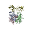
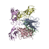
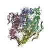
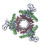
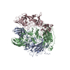
| Title | Pseudo-atomic crystal structure of the desmoglein 2 - human adenovirus serotype 3 fibre knob complex | |||||||||
 Components Components |
| |||||||||
 Keywords Keywords | VIRAL PROTEIN / virus receptor / extracellular domain / cell surface glycoprotein / desmosome | |||||||||
| Function / homology |  Function and homology information Function and homology informationPurkinje myocyte development / positive regulation of protein localization to cell-cell junction / bundle of His cell-Purkinje myocyte adhesion involved in cell communication / cell adhesive protein binding involved in bundle of His cell-Purkinje myocyte communication / desmosome organization / negative regulation of endothelial cell differentiation / Keratinization / negative regulation of inflammatory response to wounding / desmosome / mesenchymal to epithelial transition ...Purkinje myocyte development / positive regulation of protein localization to cell-cell junction / bundle of His cell-Purkinje myocyte adhesion involved in cell communication / cell adhesive protein binding involved in bundle of His cell-Purkinje myocyte communication / desmosome organization / negative regulation of endothelial cell differentiation / Keratinization / negative regulation of inflammatory response to wounding / desmosome / mesenchymal to epithelial transition / Formation of the cornified envelope / cornified envelope / regulation of ventricular cardiac muscle cell action potential / Apoptotic cleavage of cell adhesion proteins / adhesion receptor-mediated virion attachment to host cell / negative regulation of epithelial to mesenchymal transition / positive regulation of sprouting angiogenesis / homophilic cell-cell adhesion / positive regulation of stem cell population maintenance / regulation of heart rate by cardiac conduction / intercalated disc / lateral plasma membrane / RHOG GTPase cycle / RAC2 GTPase cycle / RAC3 GTPase cycle / maternal process involved in female pregnancy / cell adhesion molecule binding / positive regulation of cell adhesion / response to progesterone / stem cell proliferation / cell-cell adhesion / cell-cell junction / viral capsid / cell junction / cell adhesion / apical plasma membrane / intracellular membrane-bounded organelle / calcium ion binding / symbiont entry into host cell / negative regulation of apoptotic process / host cell nucleus / cell surface / extracellular exosome / plasma membrane / cytoplasm Similarity search - Function | |||||||||
| Biological species |  Human adenovirus B3 Human adenovirus B3 Homo sapiens (human) Homo sapiens (human) | |||||||||
| Method |  X-RAY DIFFRACTION / SYNCHROTRON / MOLECULAR REPLACEMENT / Resolution: 4.5 Å X-RAY DIFFRACTION / SYNCHROTRON / MOLECULAR REPLACEMENT / Resolution: 4.5 Å | |||||||||
 Authors Authors | Burmeister, W.P. / Fender, P. / Vassal-Stermann, E. | |||||||||
| Funding support |  France, 2items France, 2items
| |||||||||
 Citation Citation | Journal: Acta Crystallogr.,Sect.F / Year: 2019 Title: Intermediate-resolution crystal structure of the human adenovirus B serotype 3 fibre knob in complex with the EC2-EC3 fragment of desmoglein 2. Authors: Vassal-Stermann, E. / Hutin, S. / Fender, P. / Burmeister, W.P. | |||||||||
| History |
|
- Structure visualization
| Structure viewer | Molecule: MolmilJmol/JSmol |
|---|
- Downloads & links
-Download
| PDBx/mmCIF format | 6sit.cif.gz | 101 KB | Display | PDBx/mmCIF format |
|---|---|---|---|---|
| PDB format | pdb6sit.ent.gz | 75 KB | Display | PDB format |
| PDBx/mmJSON format | 6sit.json.gz | Tree view | PDBx/mmJSON format | |
| Others |  Other downloads Other downloads |
-Validation report
| Arichive directory | https://data.pdbj.org/pub/pdb/validation_reports/si/6sitftp://data.pdbj.org/pub/pdb/validation_reports/si/6sit | HTTPS FTP |
|---|
-Related structure data
| Related structure data | 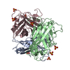 1h7zS S: Starting model for refinement |
|---|---|
| Similar structure data |


-Links
 PDBj
PDBj


- Assembly
| Deposited unit | 
| ||||||||
|---|---|---|---|---|---|---|---|---|---|
| 1 | 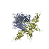
| ||||||||
| Unit cell |
|
-Components
| #1: Protein | Mass: 21270.014 Da / Num. of mol.: 1 Source method: isolated from a genetically manipulated source Source: (gene. exp.) Human adenovirus B3 / Gene: L5 / Plasmid: pETDuet-1 / Production host:  | ||
|---|---|---|---|
| #2: Protein | Mass: 26609.707 Da / Num. of mol.: 1 Source method: isolated from a genetically manipulated source Details: extracellular domain EC2 and EC3 / Source: (gene. exp.) Homo sapiens (human) / Gene: DSG2, CDHF5 / Plasmid: pETM-40 / Production host: | ||
| #3: Chemical | ChemComp-CA /   Mass: 40.078 Da / Num. of mol.: 6 / Source method: obtained synthetically / Formula: Ca / Feature type: SUBJECT OF INVESTIGATION Mass: 40.078 Da / Num. of mol.: 6 / Source method: obtained synthetically / Formula: Ca / Feature type: SUBJECT OF INVESTIGATIONHas ligand of interest | Y | |
-Experimental details
-Experiment
| Experiment | Method: X-RAY DIFFRACTION / Number of used crystals: 1 |
|---|
- Sample preparation
| Crystal | Density Matthews: 2.74 Å3/Da / Density % sol: 55.07 % / Description: prisms |
|---|---|
| Crystal grow | Temperature: 293 K / Method: vapor diffusion, hanging drop / pH: 4.6 Details: 7 mg/mL complex in 20 mM Tris-HCl, pH 8, 150 mM NaCl, 3 mM CaCl2 using a reservoir solution of 15 % PEG 3350, 0.2 M (NH4)2SO4, 0.1 M Na-acetate pH 4.6 |
-Data collection
| Diffraction | Mean temperature: 100 K / Serial crystal experiment: N | |||||||||||||||||||||||||||||||||
|---|---|---|---|---|---|---|---|---|---|---|---|---|---|---|---|---|---|---|---|---|---|---|---|---|---|---|---|---|---|---|---|---|---|---|
| Diffraction source | Source: SYNCHROTRON / Site: ESRF / Beamline: ID23-2 / Wavelength: 0.8731 Å | |||||||||||||||||||||||||||||||||
| Detector | Type: DECTRIS PILATUS3 X 2M / Detector: PIXEL / Date: Apr 12, 2018 Details: Vertically focused by CRLs and horizontally focussed by a mirror in half-Kirkpatrick-Baez (KB) geometry | |||||||||||||||||||||||||||||||||
| Radiation | Protocol: SINGLE WAVELENGTH / Monochromatic (M) / Laue (L): M / Scattering type: x-ray | |||||||||||||||||||||||||||||||||
| Radiation wavelength | Wavelength: 0.8731 Å / Relative weight: 1 | |||||||||||||||||||||||||||||||||
| Reflection | Resolution: 4.49→46.51 Å / Num. obs: 3184 / % possible obs: 97.9 % / Redundancy: 3.8 % / Biso Wilson estimate: 233 Å2 / CC1/2: 0.997 / R split: 0.093 / Rmerge(I) obs: 0.093 / Rpim(I) all: 0.053 / Rrim(I) all: 0.108 / Χ2: 0.77 / Net I/av σ(I): 4.9 / Net I/σ(I): 6.5 | |||||||||||||||||||||||||||||||||
| Reflection shell | Diffraction-ID: 1
|
- Processing
| Software |
| ||||||||||||||||||||||||
|---|---|---|---|---|---|---|---|---|---|---|---|---|---|---|---|---|---|---|---|---|---|---|---|---|---|
| Refinement | Method to determine structure: MOLECULAR REPLACEMENT Starting model: 1h7z Resolution: 4.5→46.5 Å / SU R Cruickshank DPI: 1.7 / Cross valid method: THROUGHOUT / σ(F): 0
| ||||||||||||||||||||||||
| Solvent computation | Bsol: 252.167 Å2 | ||||||||||||||||||||||||
| Displacement parameters | Biso max: 382.5 Å2 / Biso mean: 252 Å2 / Biso min: 256.54 Å2 | ||||||||||||||||||||||||
| Refinement step | Cycle: final / Resolution: 4.5→46.5 Å
| ||||||||||||||||||||||||
| Refine LS restraints |
| ||||||||||||||||||||||||
| LS refinement shell | Resolution: 4.5→4.7 Å
| ||||||||||||||||||||||||
| Xplor file |
|
