 Movie
Movie Controller


+ Open data

- Basic information
| Entry | Database: PDB / ID: 1h7z | ||||||
|---|---|---|---|---|---|---|---|
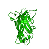
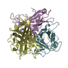
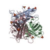
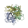
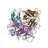
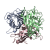
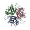
| Title | Adenovirus Ad3 fibre head | ||||||
 Components Components | ADENOVIRUS FIBRE PROTEIN | ||||||
 Keywords Keywords | CELL RECEPTOR RECOGNITION / ADENOVIRUS / AD3 / FIBRE / RECEPTOR | ||||||
| Function / homology |  Function and homology information Function and homology informationadhesion receptor-mediated virion attachment to host cell / viral capsid / cell adhesion / symbiont entry into host cell / host cell nucleus Similarity search - Function | ||||||
| Biological species |  HUMAN ADENOVIRUS TYPE 3 HUMAN ADENOVIRUS TYPE 3 | ||||||
| Method |  X-RAY DIFFRACTION / SYNCHROTRON / MOLECULAR REPLACEMENT / Resolution: 1.6 Å X-RAY DIFFRACTION / SYNCHROTRON / MOLECULAR REPLACEMENT / Resolution: 1.6 Å | ||||||
 Authors Authors | Durmort, C. / Stehlin, C. / Schoehn, G. / Mitraki, A. / Drouet, E. / Cusack, S. / Burmeister, W.P. | ||||||
 Citation Citation | Journal: Virology / Year: 2001 Title: Structure of the Fiber Head of Ad3, a Non-Car-Binding Serotype of Adenovirus Authors: Durmort, C. / Stehlin, C. / Schoehn, G. / Mitraki, A. / Drouet, E. / Cusack, S. / Burmeister, W.P. | ||||||
| History |
|
- Structure visualization
| Structure viewer | Molecule: MolmilJmol/JSmol |
|---|
- Downloads & links
-Download
| PDBx/mmCIF format | 1h7z.cif.gz | 144.8 KB | Display | PDBx/mmCIF format |
|---|---|---|---|---|
| PDB format | pdb1h7z.ent.gz | 114.2 KB | Display | PDB format |
| PDBx/mmJSON format | 1h7z.json.gz | Tree view | PDBx/mmJSON format | |
| Others |  Other downloads Other downloads |
-Validation report
| Arichive directory | https://data.pdbj.org/pub/pdb/validation_reports/h7/1h7zftp://data.pdbj.org/pub/pdb/validation_reports/h7/1h7z | HTTPS FTP |
|---|
-Related structure data
| Related structure data |  1knbS S: Starting model for refinement |
|---|---|
| Similar structure data |

-Links
 PDBj
PDBj
- Assembly
| Deposited unit | 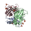
| ||||||||||||
|---|---|---|---|---|---|---|---|---|---|---|---|---|---|
| 1 |
| ||||||||||||
| Unit cell |
| ||||||||||||
| Noncrystallographic symmetry (NCS) | NCS oper:
|
-Components
| #1: Protein | Mass: 21585.447 Da / Num. of mol.: 3 / Fragment: HEAD DOMAIN RESIDUES 126-319 / Mutation: YES Source method: isolated from a genetically manipulated source Details: ONLY THE HEAD DOMAIN, START CODON INTRODUCED AT POSITION 126 Source: (gene. exp.) HUMAN ADENOVIRUS TYPE 3 / Strain: AD3 / Plasmid: PACUW31 / Cellular location (production host): CYTOPLASM / Production host:  #2: Chemical | ChemComp-SO4 /   Mass: 96.063 Da / Num. of mol.: 8 / Source method: obtained synthetically / Formula: SO4 Mass: 96.063 Da / Num. of mol.: 8 / Source method: obtained synthetically / Formula: SO4#3: Water | ChemComp-HOH / |  Mass: 18.015 Da / Num. of mol.: 891 / Source method: isolated from a natural source / Formula: H2O Mass: 18.015 Da / Num. of mol.: 891 / Source method: isolated from a natural source / Formula: H2OCompound details | CHAIN A, B, C ENGINEERED MUTATION ILE126MET SERVES AS THE LIGAND BETWEEN THE ADENOVIRUS CAPSID AND ...CHAIN A, B, C ENGINEERED | |
|---|
-Experimental details
-Experiment
| Experiment | Method: X-RAY DIFFRACTION / Number of used crystals: 1 |
|---|
- Sample preparation
| Crystal | Density Matthews: 3.51 Å3/Da / Density % sol: 64.7 % | ||||||||||||||||||||||||||||
|---|---|---|---|---|---|---|---|---|---|---|---|---|---|---|---|---|---|---|---|---|---|---|---|---|---|---|---|---|---|
| Crystal grow | pH: 7 Details: RESERVOIR: 1.5 M LI2SO4, 0.1 M HEPES PH 7.0, 7.5 MG/ML PROTEIN IN 0.02 M TRIS-HCL PH 7.0 0.001 M EDTA, 0.02 M NACL | ||||||||||||||||||||||||||||
| Crystal grow | *PLUS Method: vapor diffusion, hanging drop | ||||||||||||||||||||||||||||
| Components of the solutions | *PLUS
|
-Data collection
| Diffraction | Mean temperature: 100 K |
|---|---|
| Diffraction source | Source: SYNCHROTRON / Site: ESRF  / Beamline: ID14-1 / Wavelength: 0.933 / Beamline: ID14-1 / Wavelength: 0.933 |
| Detector | Type: MARRESEARCH / Detector: CCD / Date: Aug 15, 2000 / Details: SAGITALLY FOCUSING 2ND CRYSTAL, MULTILAYER |
| Radiation | Monochromator: DIAMOND(111) / Protocol: SINGLE WAVELENGTH / Monochromatic (M) / Laue (L): M / Scattering type: x-ray |
| Radiation wavelength | Wavelength: 0.933 Å / Relative weight: 1 |
| Reflection | Resolution: 1.6→19.389 Å / Num. obs: 108500 / % possible obs: 98.6 % / Redundancy: 3.1 % / Biso Wilson estimate: 29.4 Å2 / Rmerge(I) obs: 0.045 / Rsym value: 0.045 / Net I/σ(I): 9 |
| Reflection shell | Resolution: 1.6→1.69 Å / Redundancy: 2.8 % / Rmerge(I) obs: 0.331 / Mean I/σ(I) obs: 1.9 / Rsym value: 0.331 / % possible all: 97.9 |
| Reflection shell | *PLUS % possible obs: 97.9 % |
- Processing
| Software |
| ||||||||||||||||||||||||||||||||||||||||||||||||||||||||||||||||||||||||||||||||||||
|---|---|---|---|---|---|---|---|---|---|---|---|---|---|---|---|---|---|---|---|---|---|---|---|---|---|---|---|---|---|---|---|---|---|---|---|---|---|---|---|---|---|---|---|---|---|---|---|---|---|---|---|---|---|---|---|---|---|---|---|---|---|---|---|---|---|---|---|---|---|---|---|---|---|---|---|---|---|---|---|---|---|---|---|---|---|
| Refinement | Method to determine structure: MOLECULAR REPLACEMENT Starting model: PDB ENTRY 1KNB Resolution: 1.6→19.96 Å / SU B: 1.56 / SU ML: 0.054 / Cross valid method: THROUGHOUT / σ(F): 0 / ESU R: 0.077 / ESU R Free: 0.084 Details: RESIDUES 219 - 224 ARE POORLY DEFINED. THE CONFORMATION OF RESIDUES 240 - 246 DIFFERS FOR THE THREE MONOMERS DUE TO A CRYSTAL CONTACT IN THIS REGION THE DENSITY MAPS ONLY SHOW RESIDUES STARTING FROM 129
| ||||||||||||||||||||||||||||||||||||||||||||||||||||||||||||||||||||||||||||||||||||
| Displacement parameters | Biso mean: 36.9 Å2 | ||||||||||||||||||||||||||||||||||||||||||||||||||||||||||||||||||||||||||||||||||||
| Refinement step | Cycle: LAST / Resolution: 1.6→19.96 Å
| ||||||||||||||||||||||||||||||||||||||||||||||||||||||||||||||||||||||||||||||||||||
| Refine LS restraints |
| ||||||||||||||||||||||||||||||||||||||||||||||||||||||||||||||||||||||||||||||||||||
| Software | *PLUS Name: REFMAC / Classification: refinement | ||||||||||||||||||||||||||||||||||||||||||||||||||||||||||||||||||||||||||||||||||||
| Refinement | *PLUS Rfactor obs: 0.176 | ||||||||||||||||||||||||||||||||||||||||||||||||||||||||||||||||||||||||||||||||||||
| Solvent computation | *PLUS | ||||||||||||||||||||||||||||||||||||||||||||||||||||||||||||||||||||||||||||||||||||
| Displacement parameters | *PLUS | ||||||||||||||||||||||||||||||||||||||||||||||||||||||||||||||||||||||||||||||||||||
| LS refinement shell | *PLUS Highest resolution: 1.6 Å / Lowest resolution: 1.69 Å / Rfactor Rfree: 0.284 / Rfactor obs: 0.256 |
