 Movie
Movie Controller


+ Open data

- Basic information
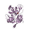
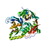
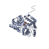
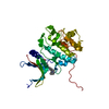
| Entry | Database: PDB / ID: 6ocf | ||||||
|---|---|---|---|---|---|---|---|
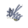
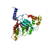
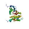
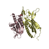
| Title | The crystal structure of VASH1-SVBP complex | ||||||
 Components Components |
| ||||||
 Keywords Keywords | HYDROLASE/PROTEIN BINDING / tubulin carboxypeptidases / microtubule modification / tubulin detyrosination / VASH1-SVBP complex / HYDROLASE-PROTEIN BINDING complex | ||||||
| Function / homology |  Function and homology information Function and homology information regulation of metallopeptidase activity / tubulinyl-Tyr carboxypeptidase / tubulin-tyrosine carboxypeptidase / Carboxyterminal post-translational modifications of tubulin / regulation of cellular senescence / negative regulation of lymphangiogenesis / peptidase activator activity / negative regulation of endothelial cell migration / labyrinthine layer blood vessel development / axon development ...regulation of metallopeptidase activity / tubulinyl-Tyr carboxypeptidase / tubulin-tyrosine carboxypeptidase / Carboxyterminal post-translational modifications of tubulin / regulation of cellular senescence / negative regulation of lymphangiogenesis / peptidase activator activity / negative regulation of endothelial cell migration / labyrinthine layer blood vessel development / axon development / negative regulation of endothelial cell proliferation / negative regulation of blood vessel endothelial cell migration / protein secretion / regulation of angiogenesis / metallocarboxypeptidase activity / negative regulation of protein ubiquitination / negative regulation of angiogenesis / response to wounding / apical part of cell / actin binding / microtubule binding / angiogenesis / cytoskeleton / regulation of cell cycle / cell cycle / endoplasmic reticulum / proteolysis / extracellular space / extracellular region / cytoplasm regulation of metallopeptidase activity / tubulinyl-Tyr carboxypeptidase / tubulin-tyrosine carboxypeptidase / Carboxyterminal post-translational modifications of tubulin / regulation of cellular senescence / negative regulation of lymphangiogenesis / peptidase activator activity / negative regulation of endothelial cell migration / labyrinthine layer blood vessel development / axon development ...regulation of metallopeptidase activity / tubulinyl-Tyr carboxypeptidase / tubulin-tyrosine carboxypeptidase / Carboxyterminal post-translational modifications of tubulin / regulation of cellular senescence / negative regulation of lymphangiogenesis / peptidase activator activity / negative regulation of endothelial cell migration / labyrinthine layer blood vessel development / axon development / negative regulation of endothelial cell proliferation / negative regulation of blood vessel endothelial cell migration / protein secretion / regulation of angiogenesis / metallocarboxypeptidase activity / negative regulation of protein ubiquitination / negative regulation of angiogenesis / response to wounding / apical part of cell / actin binding / microtubule binding / angiogenesis / cytoskeleton / regulation of cell cycle / cell cycle / endoplasmic reticulum / proteolysis / extracellular space / extracellular region / cytoplasmSimilarity search - Function | ||||||
| Biological species |  Homo sapiens (human) Homo sapiens (human) | ||||||
| Method | X-RAY DIFFRACTION / SYNCHROTRON / SAD / Resolution: 2.102 Å | ||||||
 Authors Authors | Li, F. / Luo, X. / Yu, H. | ||||||
| Funding support |  United States, 1items United States, 1items
| ||||||
 Citation Citation | Journal: Nat.Struct.Mol.Biol. / Year: 2019 Title: Structural basis of tubulin detyrosination by vasohibins. Authors: Li, F. / Hu, Y. / Qi, S. / Luo, X. / Yu, H. | ||||||
| History |
|
- Structure visualization
| Structure viewer | Molecule: MolmilJmol/JSmol |
|---|
- Downloads & links
-Download
| PDBx/mmCIF format | 6ocf.cif.gz | 143.8 KB | Display | PDBx/mmCIF format |
|---|---|---|---|---|
| PDB format | pdb6ocf.ent.gz | 117 KB | Display | PDB format |
| PDBx/mmJSON format | 6ocf.json.gz | Tree view | PDBx/mmJSON format | |
| Others |  Other downloads Other downloads |
-Validation report
| Arichive directory | https://data.pdbj.org/pub/pdb/validation_reports/oc/6ocfftp://data.pdbj.org/pub/pdb/validation_reports/oc/6ocf | HTTPS FTP |
|---|
-Related structure data


-Links
 PDBj
PDBj- Assembly
| Deposited unit | 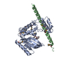
| ||||||||||
|---|---|---|---|---|---|---|---|---|---|---|---|
| 1 |
| ||||||||||
| Unit cell |
| ||||||||||
| Components on special symmetry positions |
|
-Components
| #1: Protein | / Tubulin carboxypeptidase 1 / Tyrosine carboxypeptidase 1 / TTCP 1 / Vasohibin-1 Mass: 29092.645 Da / Num. of mol.: 1 Source method: isolated from a genetically manipulated source Source: (gene. exp.) Homo sapiens (human) / Gene: VASH1, KIAA1036, VASH / Production host:  Escherichia coli BL21 (bacteria) / Strain (production host): BL21 / References: UniProt: Q7L8A9, tubulinyl-Tyr carboxypeptidase Escherichia coli BL21 (bacteria) / Strain (production host): BL21 / References: UniProt: Q7L8A9, tubulinyl-Tyr carboxypeptidase |
|---|---|
| #2: Protein/peptide | Mass: 5998.477 Da / Num. of mol.: 1 Source method: isolated from a genetically manipulated source Source: (gene. exp.) Homo sapiens (human) / Gene: SVBP, CCDC23 / Production host: Escherichia coli BL21 (bacteria) / Strain (production host): BL21 / References: UniProt: Q8N300 |
| #3: Chemical | ChemComp-GOL / Glycerol  Mass: 92.094 Da / Num. of mol.: 1 / Source method: obtained synthetically / Formula: C3H8O3 Mass: 92.094 Da / Num. of mol.: 1 / Source method: obtained synthetically / Formula: C3H8O3 |
| #4: Chemical | ChemComp-CL / Chloride  Mass: 35.453 Da / Num. of mol.: 1 / Source method: obtained synthetically / Formula: Cl Mass: 35.453 Da / Num. of mol.: 1 / Source method: obtained synthetically / Formula: Cl |
| #5: Water | ChemComp-HOH / Water Mass: 18.015 Da / Num. of mol.: 168 / Source method: isolated from a natural source / Formula: H2O Mass: 18.015 Da / Num. of mol.: 168 / Source method: isolated from a natural source / Formula: H2O |
-Experimental details
-Experiment
| Experiment | Method: X-RAY DIFFRACTION / Number of used crystals: 1 |
|---|
- Sample preparation
| Crystal | Density Matthews: 3.72 Å3/Da / Density % sol: 66.96 % |
|---|---|
| Crystal grow | Temperature: 293.15 K / Method: vapor diffusion, sitting drop / pH: 5 Details: 0.1 M sodium citrate tribasic dihydrate, pH 5.0, and 18% (w/v) PEG20000 |
-Data collection
| Diffraction | Mean temperature: 100 K / Serial crystal experiment: N | |||||||||||||||||||||||||||||||||||||||||||||||||||||||||||||||||||||||||||||||||||||||||||||||||||||||||||||||||||||||||||||||||||||||||||||||||||||||||||||||||||||||||||||||||||||||||||||
|---|---|---|---|---|---|---|---|---|---|---|---|---|---|---|---|---|---|---|---|---|---|---|---|---|---|---|---|---|---|---|---|---|---|---|---|---|---|---|---|---|---|---|---|---|---|---|---|---|---|---|---|---|---|---|---|---|---|---|---|---|---|---|---|---|---|---|---|---|---|---|---|---|---|---|---|---|---|---|---|---|---|---|---|---|---|---|---|---|---|---|---|---|---|---|---|---|---|---|---|---|---|---|---|---|---|---|---|---|---|---|---|---|---|---|---|---|---|---|---|---|---|---|---|---|---|---|---|---|---|---|---|---|---|---|---|---|---|---|---|---|---|---|---|---|---|---|---|---|---|---|---|---|---|---|---|---|---|---|---|---|---|---|---|---|---|---|---|---|---|---|---|---|---|---|---|---|---|---|---|---|---|---|---|---|---|---|---|---|---|---|
| Diffraction source | Source: SYNCHROTRON / Site: APS / Beamline: 19-ID / Wavelength: 0.9792 Å | |||||||||||||||||||||||||||||||||||||||||||||||||||||||||||||||||||||||||||||||||||||||||||||||||||||||||||||||||||||||||||||||||||||||||||||||||||||||||||||||||||||||||||||||||||||||||||||
| Detector | Type: ADSC QUANTUM 315r / Detector: CCD / Date: Mar 15, 2018 | |||||||||||||||||||||||||||||||||||||||||||||||||||||||||||||||||||||||||||||||||||||||||||||||||||||||||||||||||||||||||||||||||||||||||||||||||||||||||||||||||||||||||||||||||||||||||||||
| Radiation | Protocol: SINGLE WAVELENGTH / Monochromatic (M) / Laue (L): M / Scattering type: x-ray | |||||||||||||||||||||||||||||||||||||||||||||||||||||||||||||||||||||||||||||||||||||||||||||||||||||||||||||||||||||||||||||||||||||||||||||||||||||||||||||||||||||||||||||||||||||||||||||
| Radiation wavelength | Wavelength: 0.9792 Å / Relative weight: 1 | |||||||||||||||||||||||||||||||||||||||||||||||||||||||||||||||||||||||||||||||||||||||||||||||||||||||||||||||||||||||||||||||||||||||||||||||||||||||||||||||||||||||||||||||||||||||||||||
| Reflection | Resolution: 2.1→50 Å / Num. obs: 48368 / % possible obs: 99.6 % / Redundancy: 24.5 % / Biso Wilson estimate: 24.27 Å2 / Rmerge(I) obs: 0.088 / Rpim(I) all: 0.018 / Rrim(I) all: 0.09 / Χ2: 1.072 / Net I/σ(I): 42.8 | |||||||||||||||||||||||||||||||||||||||||||||||||||||||||||||||||||||||||||||||||||||||||||||||||||||||||||||||||||||||||||||||||||||||||||||||||||||||||||||||||||||||||||||||||||||||||||||
| Reflection shell | Diffraction-ID: 1
|
- Processing
| Software |
| ||||||||||||||||||||||||||||||||||||||||||||||||||||||||||||||||||||||||||||||||||||||||||||||||||||||||||||||||||||||||||||||||||||||||||||||||||||||
|---|---|---|---|---|---|---|---|---|---|---|---|---|---|---|---|---|---|---|---|---|---|---|---|---|---|---|---|---|---|---|---|---|---|---|---|---|---|---|---|---|---|---|---|---|---|---|---|---|---|---|---|---|---|---|---|---|---|---|---|---|---|---|---|---|---|---|---|---|---|---|---|---|---|---|---|---|---|---|---|---|---|---|---|---|---|---|---|---|---|---|---|---|---|---|---|---|---|---|---|---|---|---|---|---|---|---|---|---|---|---|---|---|---|---|---|---|---|---|---|---|---|---|---|---|---|---|---|---|---|---|---|---|---|---|---|---|---|---|---|---|---|---|---|---|---|---|---|---|---|---|---|
| Refinement | Method to determine structure: SAD / Resolution: 2.102→45.214 Å / SU ML: 0.23 / Cross valid method: THROUGHOUT / σ(F): 1.36 / Phase error: 23.71
| ||||||||||||||||||||||||||||||||||||||||||||||||||||||||||||||||||||||||||||||||||||||||||||||||||||||||||||||||||||||||||||||||||||||||||||||||||||||
| Solvent computation | Shrinkage radii: 0.9 Å / VDW probe radii: 1.11 Å | ||||||||||||||||||||||||||||||||||||||||||||||||||||||||||||||||||||||||||||||||||||||||||||||||||||||||||||||||||||||||||||||||||||||||||||||||||||||
| Displacement parameters | Biso max: 149.8 Å2 / Biso mean: 41.7347 Å2 / Biso min: 9.12 Å2 | ||||||||||||||||||||||||||||||||||||||||||||||||||||||||||||||||||||||||||||||||||||||||||||||||||||||||||||||||||||||||||||||||||||||||||||||||||||||
| Refinement step | Cycle: final / Resolution: 2.102→45.214 Å
| ||||||||||||||||||||||||||||||||||||||||||||||||||||||||||||||||||||||||||||||||||||||||||||||||||||||||||||||||||||||||||||||||||||||||||||||||||||||
| LS refinement shell | Refine-ID: X-RAY DIFFRACTION / Rfactor Rfree error: 0 / Total num. of bins used: 17
| ||||||||||||||||||||||||||||||||||||||||||||||||||||||||||||||||||||||||||||||||||||||||||||||||||||||||||||||||||||||||||||||||||||||||||||||||||||||
| Refinement TLS params. | Method: refined / Refine-ID: X-RAY DIFFRACTION
| ||||||||||||||||||||||||||||||||||||||||||||||||||||||||||||||||||||||||||||||||||||||||||||||||||||||||||||||||||||||||||||||||||||||||||||||||||||||
| Refinement TLS group |
|
