 Movie
Movie Controller


+ Open data

- Basic information
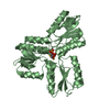
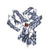
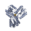
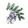
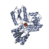
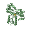
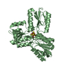
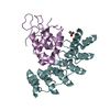
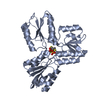
| Entry | Database: PDB / ID: 6ne4 | |||||||||
|---|---|---|---|---|---|---|---|---|---|---|
| Title | Designed repeat protein specifically in complex with Fz7CRD | |||||||||
 Components Components |
| |||||||||
 Keywords Keywords |  BIOSYNTHETIC PROTEIN/SIGNALING PROTEIN / Frizzled / Designed protein / BIOSYNTHETIC PROTEIN / BIOSYNTHETIC PROTEIN-SIGNALING PROTEIN complex BIOSYNTHETIC PROTEIN/SIGNALING PROTEIN / Frizzled / Designed protein / BIOSYNTHETIC PROTEIN / BIOSYNTHETIC PROTEIN-SIGNALING PROTEIN complex | |||||||||
| Function / homology |  Function and homology information Function and homology informationnegative regulation of ectodermal cell fate specification / negative regulation of cardiac muscle cell differentiation / mesenchymal to epithelial transition / skeletal muscle satellite cell maintenance involved in skeletal muscle regeneration / Wnt receptor activity / somatic stem cell division / non-canonical Wnt signaling pathway / Wnt-protein binding / positive regulation of epithelial cell proliferation involved in wound healing / WNT5:FZD7-mediated leishmania damping ...negative regulation of ectodermal cell fate specification / negative regulation of cardiac muscle cell differentiation / mesenchymal to epithelial transition / skeletal muscle satellite cell maintenance involved in skeletal muscle regeneration / Wnt receptor activity / somatic stem cell division / non-canonical Wnt signaling pathway / Wnt-protein binding / positive regulation of epithelial cell proliferation involved in wound healing / WNT5:FZD7-mediated leishmania damping / frizzled binding / PCP/CE pathway / regulation of canonical Wnt signaling pathway / Class B/2 (Secretin family receptors) / negative regulation of cell-substrate adhesion / Wnt signaling pathway, planar cell polarity pathway / stem cell population maintenance / canonical Wnt signaling pathway / cellular response to retinoic acid / positive regulation of phosphorylation / phosphatidylinositol-4,5-bisphosphate binding / substrate adhesion-dependent cell spreading / Asymmetric localization of PCP proteins / PDZ domain binding / G protein-coupled receptor activity / positive regulation of JNK cascade / neuron differentiation / recycling endosome membrane / T cell differentiation in thymus / positive regulation of MAPK cascade / intracellular membrane-bounded organelle / regulation of DNA-templated transcription / positive regulation of DNA-templated transcription / membrane / plasma membraneSimilarity search - Function | |||||||||
| Biological species |  Escherichia coli (E. coli) Escherichia coli (E. coli) Homo sapiens (human) Homo sapiens (human) | |||||||||
| Method | X-RAY DIFFRACTION / SYNCHROTRON / MOLECULAR REPLACEMENT / Resolution: 1.648 Å | |||||||||
 Authors Authors | Miao, Y. / Jude, K.M. / Garcia, K.C. | |||||||||
| Funding support |  United States, 2items United States, 2items
| |||||||||
 Citation Citation | Journal: Nat.Struct.Mol.Biol. / Year: 2019 Title: Receptor subtype discrimination using extensive shape complementary designed interfaces. Authors: Dang, L.T. / Miao, Y. / Ha, A. / Yuki, K. / Park, K. / Janda, C.Y. / Jude, K.M. / Mohan, K. / Ha, N. / Vallon, M. / Yuan, J. / Vilches-Moure, J.G. / Kuo, C.J. / Garcia, K.C. / Baker, D. | |||||||||
| History |
|
- Structure visualization
| Structure viewer | Molecule: MolmilJmol/JSmol |
|---|
- Downloads & links
-Download
| PDBx/mmCIF format | 6ne4.cif.gz | 139.8 KB | Display | PDBx/mmCIF format |
|---|---|---|---|---|
| PDB format | pdb6ne4.ent.gz | 108.1 KB | Display | PDB format |
| PDBx/mmJSON format | 6ne4.json.gz | Tree view | PDBx/mmJSON format | |
| Others |  Other downloads Other downloads |
-Validation report
| Arichive directory | https://data.pdbj.org/pub/pdb/validation_reports/ne/6ne4ftp://data.pdbj.org/pub/pdb/validation_reports/ne/6ne4 | HTTPS FTP |
|---|
-Related structure data
| Related structure data |  6ndzC 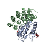 6ne1C  6ne2C  5t44S C: citing same article ( S: Starting model for refinement |
|---|---|
| Similar structure data |

-Links
 PDBj
PDBj


- Assembly
| Deposited unit | 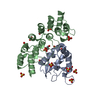
| |||||||||
|---|---|---|---|---|---|---|---|---|---|---|
| 1 |
| |||||||||
| Unit cell |
| |||||||||
| Components on special symmetry positions |
|
-Components
| #1: Protein | Mass: 20963.748 Da / Num. of mol.: 1 Source method: isolated from a genetically manipulated source Source: (gene. exp.) Escherichia coli (E. coli) / Production host: Escherichia coli (E. coli) | ||||
|---|---|---|---|---|---|
| #2: Protein | / hFz7 / FzE3 Mass: 14111.210 Da / Num. of mol.: 1 Source method: isolated from a genetically manipulated source Source: (gene. exp.) Homo sapiens (human) / Gene: FZD7 / Production host:  Trichoplusia ni (cabbage looper) / References: UniProt: O75084 Trichoplusia ni (cabbage looper) / References: UniProt: O75084 | ||||
| #3: Chemical | ChemComp-SO4 / Sulfate  Mass: 96.063 Da / Num. of mol.: 14 / Source method: obtained synthetically / Formula: SO4 Mass: 96.063 Da / Num. of mol.: 14 / Source method: obtained synthetically / Formula: SO4#4: Chemical | Ethylene glycol  Mass: 62.068 Da / Num. of mol.: 2 / Source method: obtained synthetically / Formula: C2H6O2 Mass: 62.068 Da / Num. of mol.: 2 / Source method: obtained synthetically / Formula: C2H6O2#5: Water | ChemComp-HOH / | Water Mass: 18.015 Da / Num. of mol.: 273 / Source method: isolated from a natural source / Formula: H2O Mass: 18.015 Da / Num. of mol.: 273 / Source method: isolated from a natural source / Formula: H2O |
-Experimental details
-Experiment
| Experiment | Method: X-RAY DIFFRACTION / Number of used crystals: 1 |
|---|
- Sample preparation
| Crystal | Density Matthews: 2.43 Å3/Da / Density % sol: 49.32 % |
|---|---|
| Crystal grow | Temperature: 295 K / Method: vapor diffusion, sitting drop Details: 0.2 M Na2HPO4, citric acid, pH 4.2 and 2M Ammonium sulfate |
-Data collection
| Diffraction | Mean temperature: 100 K / Serial crystal experiment: N |
|---|---|
| Diffraction source | Source: SYNCHROTRON / Site: SSRL / Beamline: BL12-2 / Wavelength: 1 Å |
| Detector | Type: DECTRIS PILATUS 6M / Detector: PIXEL / Date: Jul 23, 2017 |
| Radiation | Protocol: SINGLE WAVELENGTH / Monochromatic (M) / Laue (L): M / Scattering type: x-ray |
| Radiation wavelength | Wavelength: 1 Å / Relative weight: 1 |
| Reflection | Resolution: 1.648→50 Å / Num. obs: 41862 / % possible obs: 99.6 % / Redundancy: 11.9 % / Rmerge(I) obs: 0.084 / Net I/σ(I): 38.1 |
| Reflection shell | Resolution: 1.648→1.71 Å / Rmerge(I) obs: 0.72 / Num. unique obs: 4055 |
- Processing
| Software |
| |||||||||||||||||||||||||||||||||||||||||||||||||||||||||||||||||||||||||||||||||||||||||||||||||||||||||
|---|---|---|---|---|---|---|---|---|---|---|---|---|---|---|---|---|---|---|---|---|---|---|---|---|---|---|---|---|---|---|---|---|---|---|---|---|---|---|---|---|---|---|---|---|---|---|---|---|---|---|---|---|---|---|---|---|---|---|---|---|---|---|---|---|---|---|---|---|---|---|---|---|---|---|---|---|---|---|---|---|---|---|---|---|---|---|---|---|---|---|---|---|---|---|---|---|---|---|---|---|---|---|---|---|---|---|
| Refinement | Method to determine structure: MOLECULAR REPLACEMENT Starting model: 5T44 Resolution: 1.648→34.578 Å / SU ML: 0.17 / Cross valid method: FREE R-VALUE / σ(F): 1.38 / Phase error: 17.88
| |||||||||||||||||||||||||||||||||||||||||||||||||||||||||||||||||||||||||||||||||||||||||||||||||||||||||
| Solvent computation | Shrinkage radii: 0.9 Å / VDW probe radii: 1.11 Å | |||||||||||||||||||||||||||||||||||||||||||||||||||||||||||||||||||||||||||||||||||||||||||||||||||||||||
| Refinement step | Cycle: LAST / Resolution: 1.648→34.578 Å
| |||||||||||||||||||||||||||||||||||||||||||||||||||||||||||||||||||||||||||||||||||||||||||||||||||||||||
| Refine LS restraints |
| |||||||||||||||||||||||||||||||||||||||||||||||||||||||||||||||||||||||||||||||||||||||||||||||||||||||||
| LS refinement shell |
| |||||||||||||||||||||||||||||||||||||||||||||||||||||||||||||||||||||||||||||||||||||||||||||||||||||||||
| Refinement TLS params. | Method: refined / Refine-ID: X-RAY DIFFRACTION
| |||||||||||||||||||||||||||||||||||||||||||||||||||||||||||||||||||||||||||||||||||||||||||||||||||||||||
| Refinement TLS group |
|
