 Movie
Movie Controller

[English] 日本語
 Yorodumi
- PDB-6mos: Structure of thioredoxin 1 from the thermophilic eubacterium Ther... -
+ Open data

- Basic information
| Entry | Database: PDB / ID: 6mos | ||||||
|---|---|---|---|---|---|---|---|
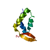
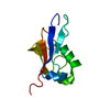
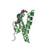
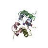
| Title | Structure of thioredoxin 1 from the thermophilic eubacterium Thermosipho africanus TCF52B | ||||||
 Components Components | Thioredoxin | ||||||
 Keywords Keywords | OXIDOREDUCTASE / thioredoxin / thermophile / Thermosipho africanus / protein stability / catalysis | ||||||
| Function / homology | Thioredoxin / Thioredoxin domain / Thioredoxin-like superfamily / Thioredoxin Function and homology information Function and homology information | ||||||
| Biological species |  Thermosipho africanus (bacteria) Thermosipho africanus (bacteria) | ||||||
| Method |  X-RAY DIFFRACTION / SYNCHROTRON / MOLECULAR REPLACEMENT / Resolution: 1.80013732031 Å X-RAY DIFFRACTION / SYNCHROTRON / MOLECULAR REPLACEMENT / Resolution: 1.80013732031 Å | ||||||
 Authors Authors | Sahtout, N. / Kuttiyatveetil, J.R. / Sanders, D.A.R. | ||||||
 Citation Citation | Journal: Biochim Biophys Acta Proteins Proteom / Year: 2019 Title: Structure and function of the putative thioredoxin 1 from the thermophilic eubacterium Thermosipho africanus strain TCF52B. Authors: Sahtout, N. / Kuttiyatveetil, J.R.A. / Sanders, D.A.R. #1: Journal: Acta Crystallogr F Struct Biol Commun / Year: 2016 Title: Putative thioredoxin Trx1 from Thermosipho africanus strain TCF52B: expression, purification and structural determination using S-SAD. Authors: Sahtout, N. / Kuttiyatveetil, J.R. / Fodje, M. / Sanders, D.A. | ||||||
| History |
|
- Structure visualization
| Structure viewer | Molecule: MolmilJmol/JSmol |
|---|
- Downloads & links
-Download
| PDBx/mmCIF format | 6mos.cif.gz | 38.5 KB | Display | PDBx/mmCIF format |
|---|---|---|---|---|
| PDB format | pdb6mos.ent.gz | 20.7 KB | Display | PDB format |
| PDBx/mmJSON format | 6mos.json.gz | Tree view | PDBx/mmJSON format | |
| Others |  Other downloads Other downloads |
-Validation report
| Summary document | 6mos_validation.pdf.gz | 434 KB | Display | wwPDB validaton report |
|---|---|---|---|---|
| Full document | 6mos_full_validation.pdf.gz | 434.4 KB | Display | |
| Data in XML | 6mos_validation.xml.gz | 6.1 KB | Display | |
| Data in CIF | 6mos_validation.cif.gz | 7.1 KB | Display | |
| Arichive directory | https://data.pdbj.org/pub/pdb/validation_reports/mo/6mosftp://data.pdbj.org/pub/pdb/validation_reports/mo/6mos | HTTPS FTP |
-Related structure data
| Similar structure data |
|---|


-Links
 PDBj
PDBj

- Assembly
| Deposited unit | 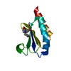
| ||||||||||||
|---|---|---|---|---|---|---|---|---|---|---|---|---|---|
| 1 |
| ||||||||||||
| Unit cell |
|
-Components
| #1: Protein | Mass: 13848.891 Da / Num. of mol.: 1 Source method: isolated from a genetically manipulated source Source: (gene. exp.) Thermosipho africanus (strain TCF52B) (bacteria)Strain: TCF52B / Gene: THA_37 / Cell line (production host): BL21-CodonPlus-RIL / Production host: |
|---|---|
| #2: Chemical | ChemComp-TAM /   Mass: 163.215 Da / Num. of mol.: 1 / Source method: obtained synthetically / Formula: C7H17NO3 / Comment: pH buffer*YM Mass: 163.215 Da / Num. of mol.: 1 / Source method: obtained synthetically / Formula: C7H17NO3 / Comment: pH buffer*YM |
| #3: Water | ChemComp-HOH /  Mass: 18.015 Da / Num. of mol.: 21 / Source method: isolated from a natural source / Formula: H2O Mass: 18.015 Da / Num. of mol.: 21 / Source method: isolated from a natural source / Formula: H2O |
-Experimental details
-Experiment
| Experiment | Method: X-RAY DIFFRACTION / Number of used crystals: 1 |
|---|
- Sample preparation
| Crystal | Density Matthews: 1.67 Å3/Da / Density % sol: 26.55 % / Description: large irregular-shaped trapezoid crystals |
|---|---|
| Crystal grow | Temperature: 289 K / Method: microbatch Details: 0.2 M MgCl2 , 0.1 M bis-tris pH 6.5, 25%(w/v) PEG 3350 |
-Data collection
| Diffraction | Mean temperature: 100 K / Serial crystal experiment: N |
|---|---|
| Diffraction source | Source: SYNCHROTRON / Site: CLSI  / Beamline: 08ID-1 / Wavelength: 0.9795 Å / Beamline: 08ID-1 / Wavelength: 0.9795 Å |
| Detector | Type: DECTRIS PILATUS3 S 6M / Detector: PIXEL / Date: Dec 4, 2015 |
| Radiation | Protocol: SINGLE WAVELENGTH / Monochromatic (M) / Laue (L): M / Scattering type: x-ray |
| Radiation wavelength | Wavelength: 0.9795 Å / Relative weight: 1 |
| Reflection | Resolution: 1.8→41.02 Å / Num. obs: 8989 / % possible obs: 99.1 % / Redundancy: 6.1 % / Biso Wilson estimate: 25.4388546055 Å2 / Net I/σ(I): 28.2 |
| Reflection shell | Resolution: 1.8→1.9 Å / Redundancy: 3.3 % / Mean I/σ(I) obs: 7.3 / Num. unique obs: 1198 / % possible all: 95 |
- Processing
| Software |
| ||||||||||||||||||||||||||||
|---|---|---|---|---|---|---|---|---|---|---|---|---|---|---|---|---|---|---|---|---|---|---|---|---|---|---|---|---|---|
| Refinement | Method to determine structure: MOLECULAR REPLACEMENT / Resolution: 1.80013732031→33.0566043115 Å / SU ML: 0.154539013252 / Cross valid method: NONE / σ(F): 1.33595773055 / Phase error: 30.8445965544
| ||||||||||||||||||||||||||||
| Solvent computation | Shrinkage radii: 0.9 Å / VDW probe radii: 1.11 Å | ||||||||||||||||||||||||||||
| Displacement parameters | Biso mean: 32.5232942362 Å2 | ||||||||||||||||||||||||||||
| Refinement step | Cycle: LAST / Resolution: 1.80013732031→33.0566043115 Å
| ||||||||||||||||||||||||||||
| Refine LS restraints |
| ||||||||||||||||||||||||||||
| LS refinement shell |
|
