 Movie
Movie Controller

[English] 日本語
 Yorodumi
- PDB-6mcj: Structure of Helical Carotenoid Protein 2 from Fremyella diplosiphon -
+ Open data

- Basic information
| Entry | Database: PDB / ID: 6mcj | ||||||
|---|---|---|---|---|---|---|---|
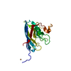
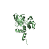
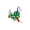
| Title | Structure of Helical Carotenoid Protein 2 from Fremyella diplosiphon | ||||||
 Components Components | Orange carotenoid-binding protein | ||||||
 Keywords Keywords | PROTEIN BINDING / PHOTOPROTECTION / CAROTENOID BINDING PROTEIN | ||||||
| Function / homology |  Function and homology information Function and homology information | ||||||
| Biological species |  Tolypothrix sp. PCC 7601 (bacteria) Tolypothrix sp. PCC 7601 (bacteria) | ||||||
| Method |  X-RAY DIFFRACTION / SYNCHROTRON / MOLECULAR REPLACEMENT / Resolution: 1.712 Å X-RAY DIFFRACTION / SYNCHROTRON / MOLECULAR REPLACEMENT / Resolution: 1.712 Å | ||||||
 Authors Authors | Sutter, M. / Dominguez-Martin, M.A. | ||||||
 Citation Citation | Journal: Biochim Biophys Acta Bioenerg / Year: 2019 Title: Structural and spectroscopic characterization of HCP2. Authors: Dominguez-Martin, M.A. / Polivka, T. / Sutter, M. / Ferlez, B. / Lechno-Yossef, S. / Montgomery, B.L. / Kerfeld, C.A. | ||||||
| History |
|
- Structure visualization
| Structure viewer | Molecule: MolmilJmol/JSmol |
|---|
- Downloads & links
-Download
| PDBx/mmCIF format | 6mcj.cif.gz | 129.2 KB | Display | PDBx/mmCIF format |
|---|---|---|---|---|
| PDB format | pdb6mcj.ent.gz | 101.6 KB | Display | PDB format |
| PDBx/mmJSON format | 6mcj.json.gz | Tree view | PDBx/mmJSON format | |
| Others |  Other downloads Other downloads |
-Validation report
| Arichive directory | https://data.pdbj.org/pub/pdb/validation_reports/mc/6mcjftp://data.pdbj.org/pub/pdb/validation_reports/mc/6mcj | HTTPS FTP |
|---|
-Related structure data
| Related structure data |  1m98 S: Starting model for refinement |
|---|---|
| Similar structure data |


-Links
 PDBj
PDBj- Assembly
| Deposited unit | 
| ||||||||
|---|---|---|---|---|---|---|---|---|---|
| 1 | 
| ||||||||
| 2 | 
| ||||||||
| Unit cell |
|
-Components
| #1: Protein | Mass: 18512.666 Da / Num. of mol.: 2 Source method: isolated from a genetically manipulated source Source: (gene. exp.) Tolypothrix sp. PCC 7601 (bacteria) / Gene: FDUTEX481_01662 / Production host: #2: Chemical |   Mass: 564.840 Da / Num. of mol.: 2 / Source method: obtained synthetically / Formula: C40H52O2 Mass: 564.840 Da / Num. of mol.: 2 / Source method: obtained synthetically / Formula: C40H52O2#3: Chemical | ChemComp-IOD /   Mass: 126.904 Da / Num. of mol.: 8 / Source method: obtained synthetically / Formula: I Mass: 126.904 Da / Num. of mol.: 8 / Source method: obtained synthetically / Formula: I#4: Chemical | ChemComp-1PE / |   Mass: 238.278 Da / Num. of mol.: 1 / Source method: obtained synthetically / Formula: C10H22O6 / Comment: precipitant*YM Mass: 238.278 Da / Num. of mol.: 1 / Source method: obtained synthetically / Formula: C10H22O6 / Comment: precipitant*YM#5: Water | ChemComp-HOH / |  Mass: 18.015 Da / Num. of mol.: 168 / Source method: isolated from a natural source / Formula: H2O Mass: 18.015 Da / Num. of mol.: 168 / Source method: isolated from a natural source / Formula: H2O |
|---|
-Experimental details
-Experiment
| Experiment | Method: X-RAY DIFFRACTION / Number of used crystals: 1 |
|---|
- Sample preparation
| Crystal | Density Matthews: 1.91 Å3/Da / Density % sol: 35.77 % |
|---|---|
| Crystal grow | Temperature: 295 K / Method: vapor diffusion, sitting drop / Details: 0.2 M Ammonium Iodide, 23 % PEG 3.350 |
-Data collection
| Diffraction | Mean temperature: 100 K |
|---|---|
| Diffraction source | Source: SYNCHROTRON / Site: ALS  / Beamline: 5.0.2 / Wavelength: 1 Å / Beamline: 5.0.2 / Wavelength: 1 Å |
| Detector | Type: DECTRIS PILATUS3 6M / Detector: PIXEL / Date: Apr 14, 2018 |
| Radiation | Protocol: SINGLE WAVELENGTH / Monochromatic (M) / Laue (L): M / Scattering type: x-ray |
| Radiation wavelength | Wavelength: 1 Å / Relative weight: 1 |
| Reflection | Resolution: 1.71→44.263 Å / Num. obs: 59576 / % possible obs: 98.8 % / Redundancy: 3.3 % / CC1/2: 0.999 / Rmerge(I) obs: 0.036 / Rpim(I) all: 0.034 / Rrim(I) all: 0.05 / Net I/σ(I): 15.7 |
| Reflection shell | Resolution: 1.71→1.8 Å / Redundancy: 3.3 % / Rmerge(I) obs: 0.273 / Mean I/σ(I) obs: 4.1 / Num. unique obs: 13974 / CC1/2: 0.893 / Rpim(I) all: 0.245 / Rrim(I) all: 0.368 / % possible all: 97.6 |
- Processing
| Software |
| ||||||||||||||||||||||||||||||||||||||||||||||||||||||||||||||||||||||||||||||||||||||||||||||||||||||||||||||||||||||||||||||||||||||||||||||||||||||||||||||||||||||||||||||||||||||||||||||||||||
|---|---|---|---|---|---|---|---|---|---|---|---|---|---|---|---|---|---|---|---|---|---|---|---|---|---|---|---|---|---|---|---|---|---|---|---|---|---|---|---|---|---|---|---|---|---|---|---|---|---|---|---|---|---|---|---|---|---|---|---|---|---|---|---|---|---|---|---|---|---|---|---|---|---|---|---|---|---|---|---|---|---|---|---|---|---|---|---|---|---|---|---|---|---|---|---|---|---|---|---|---|---|---|---|---|---|---|---|---|---|---|---|---|---|---|---|---|---|---|---|---|---|---|---|---|---|---|---|---|---|---|---|---|---|---|---|---|---|---|---|---|---|---|---|---|---|---|---|---|---|---|---|---|---|---|---|---|---|---|---|---|---|---|---|---|---|---|---|---|---|---|---|---|---|---|---|---|---|---|---|---|---|---|---|---|---|---|---|---|---|---|---|---|---|---|---|---|---|
| Refinement | Method to determine structure: MOLECULAR REPLACEMENT Starting model: 1M98 1m98 Resolution: 1.712→44.263 Å / SU ML: 0.2 / Cross valid method: FREE R-VALUE / σ(F): 1.36 / Phase error: 20
| ||||||||||||||||||||||||||||||||||||||||||||||||||||||||||||||||||||||||||||||||||||||||||||||||||||||||||||||||||||||||||||||||||||||||||||||||||||||||||||||||||||||||||||||||||||||||||||||||||||
| Solvent computation | Shrinkage radii: 0.9 Å / VDW probe radii: 1.11 Å | ||||||||||||||||||||||||||||||||||||||||||||||||||||||||||||||||||||||||||||||||||||||||||||||||||||||||||||||||||||||||||||||||||||||||||||||||||||||||||||||||||||||||||||||||||||||||||||||||||||
| Refinement step | Cycle: LAST / Resolution: 1.712→44.263 Å
| ||||||||||||||||||||||||||||||||||||||||||||||||||||||||||||||||||||||||||||||||||||||||||||||||||||||||||||||||||||||||||||||||||||||||||||||||||||||||||||||||||||||||||||||||||||||||||||||||||||
| Refine LS restraints |
| ||||||||||||||||||||||||||||||||||||||||||||||||||||||||||||||||||||||||||||||||||||||||||||||||||||||||||||||||||||||||||||||||||||||||||||||||||||||||||||||||||||||||||||||||||||||||||||||||||||
| LS refinement shell |
|
