 Movie
Movie Controller

[English] 日本語
 Yorodumi
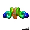
- PDB-6mam: Cleaved Ebola GP in complex with a broadly neutralizing human ant... -
+ Open data

- Basic information
| Entry | Database: PDB / ID: 6mam | |||||||||||||||
|---|---|---|---|---|---|---|---|---|---|---|---|---|---|---|---|---|
| Title | Cleaved Ebola GP in complex with a broadly neutralizing human antibody, ADI-15946 | |||||||||||||||
 Components Components |
| |||||||||||||||
 Keywords Keywords | Viral protein/immune system / Ebola / ebolavirus / antibody / mab / fab / GP / glycoprotein / cleaved / monoclonal / broad / filovirus / marburg / EBOV / BDBV / SUDV / RESTV / TAFV / MARV / RAVV / bundibugyo / sudan / human / VIRAL PROTEIN / Viral protein-immune system complex | |||||||||||||||
| Function / homology |  Function and homology information Function and homology informationsymbiont-mediated killing of host cell / host cell endoplasmic reticulum / viral budding from plasma membrane / clathrin-dependent endocytosis of virus by host cell / symbiont-mediated-mediated suppression of host tetherin activity / entry receptor-mediated virion attachment to host cell / host cell cytoplasm / symbiont-mediated suppression of host innate immune response / membrane raft / fusion of virus membrane with host endosome membrane ...symbiont-mediated killing of host cell / host cell endoplasmic reticulum / viral budding from plasma membrane / clathrin-dependent endocytosis of virus by host cell / symbiont-mediated-mediated suppression of host tetherin activity / entry receptor-mediated virion attachment to host cell / host cell cytoplasm / symbiont-mediated suppression of host innate immune response / membrane raft / fusion of virus membrane with host endosome membrane / viral envelope / symbiont entry into host cell / lipid binding / host cell plasma membrane / virion membrane / extracellular region / identical protein binding Similarity search - Function | |||||||||||||||
| Biological species |  Homo sapiens (human) Homo sapiens (human)  Zaire ebolavirus Zaire ebolavirus | |||||||||||||||
| Method |  X-RAY DIFFRACTION / SYNCHROTRON / MOLECULAR REPLACEMENT / Resolution: 4.1 Å X-RAY DIFFRACTION / SYNCHROTRON / MOLECULAR REPLACEMENT / Resolution: 4.1 Å | |||||||||||||||
 Authors Authors | West, B.R. / Moyer, C.L. / Fusco, M.L. / Saphire, E.O. | |||||||||||||||
| Funding support |  United States, 4items United States, 4items
| |||||||||||||||
 Citation Citation | Journal: Nat. Struct. Mol. Biol. / Year: 2019 Title: Structural basis of broad ebolavirus neutralization by a human survivor antibody. Authors: West, B.R. / Wec, A.Z. / Moyer, C.L. / Fusco, M.L. / Ilinykh, P.A. / Huang, K. / Wirchnianski, A.S. / James, R.M. / Herbert, A.S. / Hui, S. / Goodwin, E. / Howell, K.A. / Kailasan, S. / ...Authors: West, B.R. / Wec, A.Z. / Moyer, C.L. / Fusco, M.L. / Ilinykh, P.A. / Huang, K. / Wirchnianski, A.S. / James, R.M. / Herbert, A.S. / Hui, S. / Goodwin, E. / Howell, K.A. / Kailasan, S. / Aman, M.J. / Walker, L.M. / Dye, J.M. / Bukreyev, A. / Chandran, K. / Saphire, E.O. | |||||||||||||||
| History |
|
- Structure visualization
| Structure viewer | Molecule: MolmilJmol/JSmol |
|---|
- Downloads & links
-Download
| PDBx/mmCIF format | 6mam.cif.gz | 373 KB | Display | PDBx/mmCIF format |
|---|---|---|---|---|
| PDB format | pdb6mam.ent.gz | 306.1 KB | Display | PDB format |
| PDBx/mmJSON format | 6mam.json.gz | Tree view | PDBx/mmJSON format | |
| Others |  Other downloads Other downloads |
-Validation report
| Arichive directory | https://data.pdbj.org/pub/pdb/validation_reports/ma/6mamftp://data.pdbj.org/pub/pdb/validation_reports/ma/6mam | HTTPS FTP |
|---|
-Related structure data
| Related structure data |  5hj3S S: Starting model for refinement |
|---|---|
| Similar structure data |


-Links
 PDBj
PDBj


- Assembly
| Deposited unit | 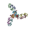
| ||||||||
|---|---|---|---|---|---|---|---|---|---|
| 1 |
| ||||||||
| Unit cell |
|
-Components
-Protein , 2 types, 6 molecules GIKHJL
| #3: Protein | Mass: 25275.326 Da / Num. of mol.: 3 Source method: isolated from a genetically manipulated source Source: (gene. exp.) Zaire ebolavirus (strain Mayinga-76) / Strain: Mayinga-76 / Gene: GP / Cell line (production host): S2 / Production host:  #4: Protein | Mass: 12254.977 Da / Num. of mol.: 3 Source method: isolated from a genetically manipulated source Source: (gene. exp.) Zaire ebolavirus (strain Mayinga-76) / Strain: Mayinga-76 / Gene: GP / Cell line (production host): S2 / Production host: |
|---|
-Antibody , 2 types, 6 molecules ACEBDF
| #1: Antibody | Mass: 25977.000 Da / Num. of mol.: 3 Source method: isolated from a genetically manipulated source Source: (gene. exp.) Homo sapiens (human) / Cell line (production host): S2 / Production host: #2: Antibody | Mass: 23793.426 Da / Num. of mol.: 3 Source method: isolated from a genetically manipulated source Source: (gene. exp.) Homo sapiens (human) / Cell line (production host): S2 / Production host: |
|---|
-Sugars , 2 types, 3 molecules
| #5: Polysaccharide | Source method: isolated from a genetically manipulated source #6: Polysaccharide | alpha-D-mannopyranose-(1-3)-beta-D-mannopyranose-(1-4)-2-acetamido-2-deoxy-beta-D-glucopyranose-(1- ...alpha-D-mannopyranose-(1-3)-beta-D-mannopyranose-(1-4)-2-acetamido-2-deoxy-beta-D-glucopyranose-(1-4)-2-acetamido-2-deoxy-beta-D-glucopyranose | Source method: isolated from a genetically manipulated source |
|---|
-Details
| Has protein modification | Y |
|---|
-Experimental details
-Experiment
| Experiment | Method: X-RAY DIFFRACTION / Number of used crystals: 1 |
|---|
- Sample preparation
| Crystal | Density Matthews: 4.15 Å3/Da / Density % sol: 70.39 % |
|---|---|
| Crystal grow | Temperature: 295 K / Method: vapor diffusion / pH: 8.2 Details: 0.2 M sodium citrate tribasic dihydrate, 20% polyethelyene glycol 3350 |
-Data collection
| Diffraction | Mean temperature: 80 K |
|---|---|
| Diffraction source | Source: SYNCHROTRON / Site: SSRL / Beamline: BL12-2 / Wavelength: 0.97946 Å |
| Detector | Type: DECTRIS PILATUS3 S 6M / Detector: PIXEL / Date: Jul 6, 2016 |
| Radiation | Protocol: SINGLE WAVELENGTH / Monochromatic (M) / Laue (L): M / Scattering type: x-ray |
| Radiation wavelength | Wavelength: 0.97946 Å / Relative weight: 1 |
| Reflection | Resolution: 4.1→48.544 Å / Num. obs: 35312 / % possible obs: 99.6 % / Redundancy: 13.1 % / CC1/2: 1 / Net I/σ(I): 14.6 |
| Reflection shell | Resolution: 4.1→4.3 Å / Redundancy: 13.4 % / Mean I/σ(I) obs: 1.8 / Num. unique obs: 3460 / CC1/2: 0.7 / % possible all: 100 |
- Processing
| Software |
| ||||||||||||||||||||||||||||||||||||||||||||||||||||||||||||||||||||||||||||||||||||||||||||||||||
|---|---|---|---|---|---|---|---|---|---|---|---|---|---|---|---|---|---|---|---|---|---|---|---|---|---|---|---|---|---|---|---|---|---|---|---|---|---|---|---|---|---|---|---|---|---|---|---|---|---|---|---|---|---|---|---|---|---|---|---|---|---|---|---|---|---|---|---|---|---|---|---|---|---|---|---|---|---|---|---|---|---|---|---|---|---|---|---|---|---|---|---|---|---|---|---|---|---|---|---|
| Refinement | Method to determine structure: MOLECULAR REPLACEMENT Starting model: 5hj3 Resolution: 4.1→48.544 Å / SU ML: 0.54 / Cross valid method: FREE R-VALUE / σ(F): 1.33 / Phase error: 29.34
| ||||||||||||||||||||||||||||||||||||||||||||||||||||||||||||||||||||||||||||||||||||||||||||||||||
| Solvent computation | Shrinkage radii: 0.9 Å / VDW probe radii: 1.11 Å | ||||||||||||||||||||||||||||||||||||||||||||||||||||||||||||||||||||||||||||||||||||||||||||||||||
| Refinement step | Cycle: LAST / Resolution: 4.1→48.544 Å
| ||||||||||||||||||||||||||||||||||||||||||||||||||||||||||||||||||||||||||||||||||||||||||||||||||
| Refine LS restraints |
| ||||||||||||||||||||||||||||||||||||||||||||||||||||||||||||||||||||||||||||||||||||||||||||||||||
| LS refinement shell |
|
