 Movie
Movie Controller

[English] 日本語
 Yorodumi
- PDB-6l9t: Crystal structure of the complex of bovine lactoperoxidase with O... -
+ Open data

- Basic information
| Entry | Database: PDB / ID: 6l9t | |||||||||
|---|---|---|---|---|---|---|---|---|---|---|
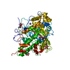
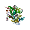
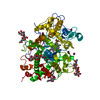
| Title | Crystal structure of the complex of bovine lactoperoxidase with OSCN at 1.89 A resolution | |||||||||
 Components Components | Lactoperoxidase | |||||||||
 Keywords Keywords | OXIDOREDUCTASE / lactoperoxidase | |||||||||
| Function / homology |  Function and homology information Function and homology informationEvents associated with phagocytolytic activity of PMN cells / thiocyanate peroxidase activity / lactoperoxidase activity / peroxidase / hydrogen peroxide catabolic process / peroxidase activity / antibacterial humoral response / response to oxidative stress / defense response to bacterium / calcium ion binding ...Events associated with phagocytolytic activity of PMN cells / thiocyanate peroxidase activity / lactoperoxidase activity / peroxidase / hydrogen peroxide catabolic process / peroxidase activity / antibacterial humoral response / response to oxidative stress / defense response to bacterium / calcium ion binding / heme binding / extracellular space / cytoplasmSimilarity search - Function | |||||||||
| Biological species |  Bos taurus (cattle) Bos taurus (cattle) | |||||||||
| Method | X-RAY DIFFRACTION / SYNCHROTRON / MOLECULAR REPLACEMENT / Resolution: 1.89 Å | |||||||||
 Authors Authors | Singh, P.K. / Viswanathan, V. / Pandey, N. / Singh, A. / Sinha, M. / Singh, R.P. / Kaur, P. / Sharma, S. / Singh, T.P. | |||||||||
 Citation Citation | Journal: To Be Published Title: Crystal structure of the complex of bovine lactoperoxidase with OSCN at 1.89 A resolution Authors: Singh, P.K. / Kaur, P. / Sharma, S. / Singh, T.P. | |||||||||
| History |
|
- Structure visualization
| Structure viewer | Molecule: MolmilJmol/JSmol |
|---|
- Downloads & links
-Download
| PDBx/mmCIF format | 6l9t.cif.gz | 161.3 KB | Display | PDBx/mmCIF format |
|---|---|---|---|---|
| PDB format | pdb6l9t.ent.gz | Display | PDB format | |
| PDBx/mmJSON format | 6l9t.json.gz | Tree view | PDBx/mmJSON format | |
| Others |  Other downloads Other downloads |
-Validation report
| Arichive directory | https://data.pdbj.org/pub/pdb/validation_reports/l9/6l9tftp://data.pdbj.org/pub/pdb/validation_reports/l9/6l9t | HTTPS FTP |
|---|
-Related structure data
| Related structure data |  3ogw S: Starting model for refinement |
|---|---|
| Similar structure data |


-Links
 PDBj
PDBj

- Assembly
| Deposited unit | 
| ||||||||
|---|---|---|---|---|---|---|---|---|---|
| 1 |
| ||||||||
| Unit cell |
|
-Components
-Protein , 1 types, 1 molecules A
| #1: Protein | / LPO Mass: 67773.305 Da / Num. of mol.: 1 / Source method: isolated from a natural source / Source: (natural) Bos taurus (cattle) / References: UniProt: P80025, peroxidase |
|---|
-Sugars , 2 types, 4 molecules 
| #2: Polysaccharide | / Mass: 424.401 Da / Num. of mol.: 2 Source method: isolated from a genetically manipulated source #6: Sugar | N-Acetylglucosamine Type: D-saccharide, beta linking / Mass: 221.208 Da / Num. of mol.: 2 / Source method: obtained synthetically / Formula: C8H15NO6 / Feature type: SUBJECT OF INVESTIGATION Type: D-saccharide, beta linking / Mass: 221.208 Da / Num. of mol.: 2 / Source method: obtained synthetically / Formula: C8H15NO6 / Feature type: SUBJECT OF INVESTIGATION |
|---|
-Non-polymers , 7 types, 594 molecules 


| #3: Chemical | ChemComp-CA /  Mass: 40.078 Da / Num. of mol.: 1 / Source method: obtained synthetically / Formula: Ca Mass: 40.078 Da / Num. of mol.: 1 / Source method: obtained synthetically / Formula: Ca | ||||||
|---|---|---|---|---|---|---|---|
| #4: Chemical | ChemComp-ZN /  Mass: 65.409 Da / Num. of mol.: 1 / Source method: obtained synthetically / Formula: Zn Mass: 65.409 Da / Num. of mol.: 1 / Source method: obtained synthetically / Formula: Zn | ||||||
| #5: Chemical | ChemComp-HEM / Heme B Mass: 616.487 Da / Num. of mol.: 1 / Source method: obtained synthetically / Formula: C34H32FeN4O4 / Feature type: SUBJECT OF INVESTIGATION Mass: 616.487 Da / Num. of mol.: 1 / Source method: obtained synthetically / Formula: C34H32FeN4O4 / Feature type: SUBJECT OF INVESTIGATION | ||||||
| #7: Chemical | ChemComp-IOD / Iodide Mass: 126.904 Da / Num. of mol.: 19 Mass: 126.904 Da / Num. of mol.: 19Source method: isolated from a genetically manipulated source Formula: I #8: Chemical | ChemComp-SCN / Thiocyanate Mass: 58.082 Da / Num. of mol.: 9 / Source method: obtained synthetically / Formula: CNS Mass: 58.082 Da / Num. of mol.: 9 / Source method: obtained synthetically / Formula: CNS#9: Chemical | ChemComp-OSM / |  Mass: 79.122 Da / Num. of mol.: 1 / Source method: obtained synthetically / Formula: CH5NOS / Feature type: SUBJECT OF INVESTIGATION Mass: 79.122 Da / Num. of mol.: 1 / Source method: obtained synthetically / Formula: CH5NOS / Feature type: SUBJECT OF INVESTIGATION#10: Water | ChemComp-HOH / | WaterMass: 18.015 Da / Num. of mol.: 562 / Source method: isolated from a natural source / Formula: H2O |
-Details
| Has ligand of interest | Y |
|---|
-Experimental details
-Experiment
| Experiment | Method: X-RAY DIFFRACTION / Number of used crystals: 1 |
|---|
- Sample preparation
| Crystal | Density Matthews: 2.37 Å3/Da / Density % sol: 48.14 % |
|---|---|
| Crystal grow | Temperature: 298 K / Method: vapor diffusion, hanging drop / pH: 6.8 Details: Ammonium Iodide, PEG3350, pH6.8, VAPOR DIFFUSION, HANGING DROP, temperature 298K PH range: 5-8 |
-Data collection
| Diffraction | Mean temperature: 100 K / Serial crystal experiment: N |
|---|---|
| Diffraction source | Source: SYNCHROTRON / Site: ESRF  / Beamline: BM14 / Wavelength: 0.97 Å / Beamline: BM14 / Wavelength: 0.97 Å |
| Detector | Type: MARRESEARCH / Detector: CCD / Date: May 13, 2010 / Details: mirror |
| Radiation | Monochromator: graphite / Protocol: SINGLE WAVELENGTH / Monochromatic (M) / Laue (L): M / Scattering type: x-ray |
| Radiation wavelength | Wavelength: 0.97 Å / Relative weight: 1 |
| Reflection | Resolution: 1.89→74.59 Å / Num. obs: 50831 / % possible obs: 99.4 % / Redundancy: 8.2 % / Rsym value: 0.091 / Net I/σ(I): 10.8 |
| Reflection shell | Resolution: 1.89→1.94 Å / Num. unique obs: 3581 / Rsym value: 0.341 / % possible all: 100 |
- Processing
| Software |
| ||||||||||||||||||||||||||||||||||||||||||||||||||||||||||||||||||||||||||||||||||||||||||||||||||||||||||||||||||||||||||||||||||||||||||||||||||||||||||||||||||||||||||
|---|---|---|---|---|---|---|---|---|---|---|---|---|---|---|---|---|---|---|---|---|---|---|---|---|---|---|---|---|---|---|---|---|---|---|---|---|---|---|---|---|---|---|---|---|---|---|---|---|---|---|---|---|---|---|---|---|---|---|---|---|---|---|---|---|---|---|---|---|---|---|---|---|---|---|---|---|---|---|---|---|---|---|---|---|---|---|---|---|---|---|---|---|---|---|---|---|---|---|---|---|---|---|---|---|---|---|---|---|---|---|---|---|---|---|---|---|---|---|---|---|---|---|---|---|---|---|---|---|---|---|---|---|---|---|---|---|---|---|---|---|---|---|---|---|---|---|---|---|---|---|---|---|---|---|---|---|---|---|---|---|---|---|---|---|---|---|---|---|---|---|---|
| Refinement | Method to determine structure: MOLECULAR REPLACEMENT Starting model: 3ogw 3ogw Resolution: 1.89→35.325 Å / Cor.coef. Fo:Fc: 0.948 / Cor.coef. Fo:Fc free: 0.913 / WRfactor Rfree: 0.278 / WRfactor Rwork: 0.217 / SU B: 4.554 / SU ML: 0.135 / Average fsc free: 0.8773 / Average fsc work: 0.9028 / Cross valid method: FREE R-VALUE / ESU R: 0.181 / ESU R Free: 0.172 Details: Hydrogens have been added in their riding positions
| ||||||||||||||||||||||||||||||||||||||||||||||||||||||||||||||||||||||||||||||||||||||||||||||||||||||||||||||||||||||||||||||||||||||||||||||||||||||||||||||||||||||||||
| Solvent computation | Ion probe radii: 0.8 Å / Shrinkage radii: 0.8 Å / VDW probe radii: 1.2 Å / Solvent model: MASK BULK SOLVENT | ||||||||||||||||||||||||||||||||||||||||||||||||||||||||||||||||||||||||||||||||||||||||||||||||||||||||||||||||||||||||||||||||||||||||||||||||||||||||||||||||||||||||||
| Displacement parameters | Biso mean: 33.677 Å2
| ||||||||||||||||||||||||||||||||||||||||||||||||||||||||||||||||||||||||||||||||||||||||||||||||||||||||||||||||||||||||||||||||||||||||||||||||||||||||||||||||||||||||||
| Refinement step | Cycle: LAST / Resolution: 1.89→35.325 Å
| ||||||||||||||||||||||||||||||||||||||||||||||||||||||||||||||||||||||||||||||||||||||||||||||||||||||||||||||||||||||||||||||||||||||||||||||||||||||||||||||||||||||||||
| Refine LS restraints |
| ||||||||||||||||||||||||||||||||||||||||||||||||||||||||||||||||||||||||||||||||||||||||||||||||||||||||||||||||||||||||||||||||||||||||||||||||||||||||||||||||||||||||||
| LS refinement shell |
|
