Resolution: 2.3→54.46 Å / Cor.coef. Fo:Fc: 0.852 / Cor.coef. Fo:Fc free: 0.82 / SU B: 10.255 / SU ML: 0.251 / Cross valid method: THROUGHOUT / ESU R: 0.542 / ESU R Free: 0.306 Details: Hydrogens have been added in their riding positions
Rfactor
Num. reflection
% reflection
Rfree
0.2951
1401
5.04 %
Rwork
0.2454
-
-
all
0.248
-
-
obs
-
27800
98.725 %
Solvent computation
Ion probe radii: 0.8 Å / Shrinkage radii: 0.8 Å / VDW probe radii: 1.2 Å
Displacement parameters
Biso mean: 40.537 Å2
Baniso -1
Baniso -2
Baniso -3
1-
0.617 Å2
-0 Å2
-1.481 Å2
2-
-
-1.246 Å2
-0 Å2
3-
-
-
1.174 Å2
Refinement step
Cycle: LAST / Resolution: 2.3→54.46 Å
Protein
Nucleic acid
Ligand
Solvent
Total
Num. atoms
4774
0
119
251
5144
Refine LS restraints
Refine-ID
Type
Dev ideal
Dev ideal target
Number
X-RAY DIFFRACTION
r_bond_refined_d
0.007
0.013
5029
X-RAY DIFFRACTION
r_bond_other_d
0.002
0.017
4558
X-RAY DIFFRACTION
r_angle_refined_deg
1.51
1.68
6836
X-RAY DIFFRACTION
r_angle_other_deg
1.206
1.598
10590
X-RAY DIFFRACTION
r_dihedral_angle_1_deg
7.863
5
594
X-RAY DIFFRACTION
r_dihedral_angle_2_deg
34.962
21.841
277
X-RAY DIFFRACTION
r_dihedral_angle_3_deg
16.741
15
824
X-RAY DIFFRACTION
r_dihedral_angle_4_deg
14.093
15
38
X-RAY DIFFRACTION
r_chiral_restr
0.068
0.2
625
X-RAY DIFFRACTION
r_gen_planes_refined
0.006
0.02
5608
X-RAY DIFFRACTION
r_gen_planes_other
0.003
0.02
1099
X-RAY DIFFRACTION
r_nbd_refined
0.221
0.2
1254
X-RAY DIFFRACTION
r_symmetry_nbd_other
0.197
0.2
4804
X-RAY DIFFRACTION
r_nbtor_refined
0.166
0.2
2366
X-RAY DIFFRACTION
r_symmetry_nbtor_other
0.078
0.2
2167
X-RAY DIFFRACTION
r_xyhbond_nbd_refined
0.206
0.2
273
X-RAY DIFFRACTION
r_symmetry_xyhbond_nbd_other
0.123
0.2
5
X-RAY DIFFRACTION
r_symmetry_nbd_refined
0.269
0.2
25
X-RAY DIFFRACTION
r_nbd_other
0.299
0.2
58
X-RAY DIFFRACTION
r_symmetry_xyhbond_nbd_refined
0.27
0.2
7
X-RAY DIFFRACTION
r_xyhbond_nbd_other
0.075
0.2
1
X-RAY DIFFRACTION
r_symmetry_metal_ion_refined
0.736
0.2
1
X-RAY DIFFRACTION
r_mcbond_it
2.998
4.31
2382
X-RAY DIFFRACTION
r_mcbond_other
2.993
4.307
2381
X-RAY DIFFRACTION
r_mcangle_it
4.7
6.455
2972
X-RAY DIFFRACTION
r_mcangle_other
4.7
6.458
2973
X-RAY DIFFRACTION
r_scbond_it
2.763
4.474
2647
X-RAY DIFFRACTION
r_scbond_other
2.761
4.474
2647
X-RAY DIFFRACTION
r_scangle_it
4.328
6.652
3864
X-RAY DIFFRACTION
r_scangle_other
4.328
6.652
3865
X-RAY DIFFRACTION
r_lrange_it
7.099
49.226
5894
X-RAY DIFFRACTION
r_lrange_other
7.101
49.225
5893
LS refinement shell
Resolution (Å)
Rfactor Rfree
Num. reflection Rfree
Rfactor Rwork
Num. reflection Rwork
Refine-ID
% reflection obs (%)
2.3-2.36
0.317
101
0.19
1999
X-RAY DIFFRACTION
100
2.36-2.424
0.292
96
0.19
1883
X-RAY DIFFRACTION
100
2.424-2.495
0.285
95
0.19
1875
X-RAY DIFFRACTION
100
2.495-2.571
0.315
109
0.19
1792
X-RAY DIFFRACTION
99.9474
2.571-2.655
0.249
92
0.19
1738
X-RAY DIFFRACTION
99.9454
2.655-2.748
0.334
90
0.19
1714
X-RAY DIFFRACTION
99.7788
2.748-2.852
0.305
80
0.19
1649
X-RAY DIFFRACTION
99.9422
2.852-2.968
0.304
73
0.19
1591
X-RAY DIFFRACTION
100
2.968-3.1
0.273
102
0.19
1478
X-RAY DIFFRACTION
99.9367
3.1-3.251
0.26
86
0.19
1440
X-RAY DIFFRACTION
99.9345
3.251-3.427
0.346
62
0.19
1350
X-RAY DIFFRACTION
96.4481
3.427-3.634
0.424
59
0.19
1269
X-RAY DIFFRACTION
96.6521
3.634-3.884
0.27
54
0.19
1118
X-RAY DIFFRACTION
90.8527
3.884-4.195
0.231
58
0.19
1037
X-RAY DIFFRACTION
89.3878
4.195-4.593
0.193
57
0.159
1048
X-RAY DIFFRACTION
100
4.593-5.133
0.202
66
0.152
941
X-RAY DIFFRACTION
100
5.133-5.922
0.336
44
0.191
860
X-RAY DIFFRACTION
100
5.922-7.242
0.219
38
0.19
711
X-RAY DIFFRACTION
100
7.242-10
0.225
29
0.168
573
X-RAY DIFFRACTION
100
8-10
0.219
10
0.19
333
X-RAY DIFFRACTION
99.1329
+
About Yorodumi
-
News
-
Feb 9, 2022. New format data for meta-information of EMDB entries
New format data for meta-information of EMDB entries
Version 3 of the EMDB header file is now the official format.
The previous official version 1.9 will be removed from the archive.
In the structure databanks used in Yorodumi, some data are registered as the other names, "COVID-19 virus" and "2019-nCoV". Here are the details of the virus and the list of structure data.
Jan 31, 2019. EMDB accession codes are about to change! (news from PDBe EMDB page)
EMDB accession codes are about to change! (news from PDBe EMDB page)
The allocation of 4 digits for EMDB accession codes will soon come to an end. Whilst these codes will remain in use, new EMDB accession codes will include an additional digit and will expand incrementally as the available range of codes is exhausted. The current 4-digit format prefixed with “EMD-” (i.e. EMD-XXXX) will advance to a 5-digit format (i.e. EMD-XXXXX), and so on. It is currently estimated that the 4-digit codes will be depleted around Spring 2019, at which point the 5-digit format will come into force.
The EM Navigator/Yorodumi systems omit the EMD- prefix.
Related info.:Q: What is EMD? / ID/Accession-code notation in Yorodumi/EM Navigator
Yorodumi is a browser for structure data from EMDB, PDB, SASBDB, etc.
This page is also the successor to EM Navigator detail page, and also detail information page/front-end page for Omokage search.
The word "yorodu" (or yorozu) is an old Japanese word meaning "ten thousand". "mi" (miru) is to see.
Related info.:EMDB / PDB / SASBDB / Comparison of 3 databanks / Yorodumi Search / Aug 31, 2016. New EM Navigator & Yorodumi / Yorodumi Papers / Jmol/JSmol / Function and homology information / Changes in new EM Navigator and Yorodumi
 Movie
Movie Controller

 Yorodumi
 Open data
 Basic information
 Components
 Keywords
 Function and homology information
 X-RAY DIFFRACTION /
  Authors
 Citation
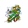
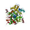
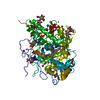
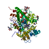
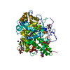
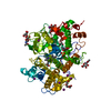
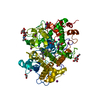
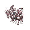
 Structure visualization
 Downloads & links
 Other downloads


 PDBj
PDBj
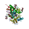
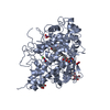
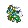
 Assembly

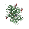
 Type: D-saccharide, beta linking / Mass: 221.208 Da / Num. of mol.: 4


 Mass: 616.487 Da / Num. of mol.: 1 / Source method: obtained synthetically / Formula: C34H32FeN4O4 / Feature type: SUBJECT OF INVESTIGATION
 Mass: 40.078 Da / Num. of mol.: 1 / Source method: obtained synthetically / Formula: Ca
 Mass: 126.904 Da / Num. of mol.: 15 / Source method: obtained synthetically / Formula: I
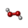
 Mass: 34.015 Da / Num. of mol.: 2 / Source method: obtained synthetically / Formula: H2O2
 Sample preparation
 / Beamline: PX-BL21 / Wavelength: 0.9795 Å
 Processing