 Movie
Movie Controller


+ Open data

- Basic information
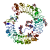
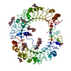
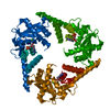
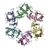
| Entry | Database: PDB / ID: 6hrr | ||||||
|---|---|---|---|---|---|---|---|
| Title | Structure of the TRPML2 ELD at pH 6.5 | ||||||
 Components Components | Mucolipin-2 | ||||||
 Keywords Keywords | MEMBRANE PROTEIN / TRPML Channel Mucolipin Cation Channel Endolysosomal System | ||||||
| Function / homology |  Function and homology information Function and homology informationpositive regulation of macrophage inflammatory protein 1 alpha production / macrophage migration / NAADP-sensitive calcium-release channel activity / positive regulation of chemokine (C-C motif) ligand 5 production / iron ion transmembrane transporter activity / neutrophil migration / positive regulation of monocyte chemotactic protein-1 production / positive regulation of chemokine (C-X-C motif) ligand 2 production / TRP channels / calcium ion transmembrane transport ...positive regulation of macrophage inflammatory protein 1 alpha production / macrophage migration / NAADP-sensitive calcium-release channel activity / positive regulation of chemokine (C-C motif) ligand 5 production / iron ion transmembrane transporter activity / neutrophil migration / positive regulation of monocyte chemotactic protein-1 production / positive regulation of chemokine (C-X-C motif) ligand 2 production / TRP channels / calcium ion transmembrane transport / calcium channel activity / recycling endosome membrane / late endosome membrane / protein transport / adaptive immune response / lysosome / innate immune response / identical protein binding / membrane / plasma membrane Similarity search - Function | ||||||
| Biological species |  Homo sapiens (human) Homo sapiens (human) | ||||||
| Method |  X-RAY DIFFRACTION / SYNCHROTRON / MOLECULAR REPLACEMENT / molecular replacement / Resolution: 2 Å X-RAY DIFFRACTION / SYNCHROTRON / MOLECULAR REPLACEMENT / molecular replacement / Resolution: 2 Å | ||||||
 Authors Authors | Bader, N. / Viet, K.K. / Wagner, A. / Hellmich, U.A. | ||||||
 Citation Citation | Journal: Structure / Year: 2019 Title: Structure of the Human TRPML2 Ion Channel Extracytosolic/Lumenal Domain. Authors: Kerstin K Viet / Annika Wagner / Kevin Schwickert / Nils Hellwig / Martha Brennich / Nicole Bader / Tanja Schirmeister / Nina Morgner / Hermann Schindelin / Ute A Hellmich /   Abstract: TRPML2 is the least structurally characterized mammalian transient receptor potential mucolipin ion channel. The TRPML family hallmark is a large extracytosolic/lumenal domain (ELD) between ...TRPML2 is the least structurally characterized mammalian transient receptor potential mucolipin ion channel. The TRPML family hallmark is a large extracytosolic/lumenal domain (ELD) between transmembrane helices S1 and S2. We present crystal structures of the tetrameric human TRPML2 ELD at pH 6.5 (2.0 Å) and 4.5 (2.95 Å), corresponding to the pH values in recycling endosomes and lysosomes. Isothermal titration calorimetry shows Ca binding to the highly acidic central pre-pore loop which is abrogated at low pH, in line with a pH-dependent channel regulation model. Small angle X-ray scattering confirms the ELD dimensions in solution. Changes in pH or Ca concentration do not affect the protein's secondary structure, but can influence ELD oligomer integrity according to native mass spectrometry. Our data thus complete the set of high-resolution views of human TRPML channel ELDs and reveal some structural responses to the conditions the TRPML2 ELD encounters as the channel traffics through the endolysosomal system. | ||||||
| History |
|
- Structure visualization
| Structure viewer | Molecule: MolmilJmol/JSmol |
|---|
- Downloads & links
-Download
| PDBx/mmCIF format | 6hrr.cif.gz | 303.3 KB | Display | PDBx/mmCIF format |
|---|---|---|---|---|
| PDB format | pdb6hrr.ent.gz | 250.1 KB | Display | PDB format |
| PDBx/mmJSON format | 6hrr.json.gz | Tree view | PDBx/mmJSON format | |
| Others |  Other downloads Other downloads |
-Validation report
| Arichive directory | https://data.pdbj.org/pub/pdb/validation_reports/hr/6hrrftp://data.pdbj.org/pub/pdb/validation_reports/hr/6hrr | HTTPS FTP |
|---|
-Related structure data
| Related structure data |  6hrsC 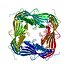 5tjaS S: Starting model for refinement C: citing same article ( |
|---|---|
| Similar structure data |


-Links
 PDBj
PDBj- Assembly
| Deposited unit | 
| ||||||||
|---|---|---|---|---|---|---|---|---|---|
| 1 | 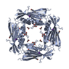
| ||||||||
| 2 | 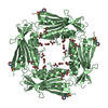
| ||||||||
| Unit cell |
| ||||||||
| Components on special symmetry positions |
|
-Components
-Protein , 1 types, 2 molecules AB
| #1: Protein | Mass: 22212.836 Da / Num. of mol.: 2 Source method: isolated from a genetically manipulated source Source: (gene. exp.) Homo sapiens (human) / Gene: MCOLN2 / Production host:  |
|---|
-Non-polymers , 5 types, 224 molecules 


| #2: Chemical | ChemComp-K /  Mass: 39.098 Da / Num. of mol.: 1 / Source method: obtained synthetically / Formula: K Mass: 39.098 Da / Num. of mol.: 1 / Source method: obtained synthetically / Formula: K | ||||||
|---|---|---|---|---|---|---|---|
| #3: Chemical |  Mass: 35.453 Da / Num. of mol.: 3 / Source method: obtained synthetically / Formula: Cl Mass: 35.453 Da / Num. of mol.: 3 / Source method: obtained synthetically / Formula: Cl#4: Chemical | ChemComp-GOL /  Mass: 92.094 Da / Num. of mol.: 11 / Source method: obtained synthetically / Formula: C3H8O3 Mass: 92.094 Da / Num. of mol.: 11 / Source method: obtained synthetically / Formula: C3H8O3#5: Chemical |  Mass: 195.237 Da / Num. of mol.: 3 / Source method: obtained synthetically / Formula: C6H13NO4S / Comment: pH buffer*YM Mass: 195.237 Da / Num. of mol.: 3 / Source method: obtained synthetically / Formula: C6H13NO4S / Comment: pH buffer*YM#6: Water | ChemComp-HOH / | Mass: 18.015 Da / Num. of mol.: 206 / Source method: isolated from a natural source / Formula: H2O |
-Details
| Has protein modification | Y |
|---|
-Experimental details
-Experiment
| Experiment | Method: X-RAY DIFFRACTION / Number of used crystals: 1 |
|---|
- Sample preparation
| Crystal | Density Matthews: 2.52 Å3/Da / Density % sol: 51.28 % / Description: Cube |
|---|---|
| Crystal grow | Temperature: 293 K / Method: vapor diffusion, hanging drop / pH: 6.5 / Details: 30% Jeffamine M-600 0.1 M MES pH 6.5 50 mM CsCl |
-Data collection
| Diffraction | Mean temperature: 100 K |
|---|---|
| Diffraction source | Source: SYNCHROTRON / Site: ESRF / Beamline: ID30B / Wavelength: 0.9677 Å |
| Detector | Type: DECTRIS PILATUS3 S 6M / Detector: PIXEL / Date: May 11, 2018 |
| Radiation | Protocol: SINGLE WAVELENGTH / Monochromatic (M) / Laue (L): M / Scattering type: x-ray |
| Radiation wavelength | Wavelength: 0.9677 Å / Relative weight: 1 |
| Reflection | Resolution: 2→46.53 Å / Num. obs: 31030 / % possible obs: 99.9 % / Redundancy: 27.2 % / Biso Wilson estimate: 47.23 Å2 / CC1/2: 1 / Rmerge(I) obs: 0.102 / Rpim(I) all: 0.002 / Net I/σ(I): 21.5 |
| Reflection shell | Resolution: 2→2.05 Å / Redundancy: 28.8 % / Rmerge(I) obs: 3.556 / Mean I/σ(I) obs: 1.3 / Num. unique obs: 2240 / CC1/2: 0.645 / Rpim(I) all: 0.673 / % possible all: 100 |
-Phasing
| Phasing | Method: molecular replacement |
|---|
- Processing
| Software |
| ||||||||||||||||||||||||||||||||||||||||||||||||||||||||||||||||||||||||||||||||||||||||||||||||||||||||||||
|---|---|---|---|---|---|---|---|---|---|---|---|---|---|---|---|---|---|---|---|---|---|---|---|---|---|---|---|---|---|---|---|---|---|---|---|---|---|---|---|---|---|---|---|---|---|---|---|---|---|---|---|---|---|---|---|---|---|---|---|---|---|---|---|---|---|---|---|---|---|---|---|---|---|---|---|---|---|---|---|---|---|---|---|---|---|---|---|---|---|---|---|---|---|---|---|---|---|---|---|---|---|---|---|---|---|---|---|---|---|
| Refinement | Method to determine structure: MOLECULAR REPLACEMENT Starting model: 5TJA Resolution: 2→20 Å / Cor.coef. Fo:Fc: 0.952 / Cor.coef. Fo:Fc free: 0.943 / SU R Cruickshank DPI: 0.263 / Cross valid method: THROUGHOUT / σ(F): 0 / SU R Blow DPI: 0.166 / SU Rfree Blow DPI: 0.143 / SU Rfree Cruickshank DPI: 0.141
| ||||||||||||||||||||||||||||||||||||||||||||||||||||||||||||||||||||||||||||||||||||||||||||||||||||||||||||
| Displacement parameters | Biso max: 202.68 Å2 / Biso mean: 61.21 Å2 / Biso min: 23.95 Å2
| ||||||||||||||||||||||||||||||||||||||||||||||||||||||||||||||||||||||||||||||||||||||||||||||||||||||||||||
| Refine analyze | Luzzati coordinate error obs: 0.24 Å | ||||||||||||||||||||||||||||||||||||||||||||||||||||||||||||||||||||||||||||||||||||||||||||||||||||||||||||
| Refinement step | Cycle: final / Resolution: 2→20 Å
| ||||||||||||||||||||||||||||||||||||||||||||||||||||||||||||||||||||||||||||||||||||||||||||||||||||||||||||
| Refine LS restraints |
| ||||||||||||||||||||||||||||||||||||||||||||||||||||||||||||||||||||||||||||||||||||||||||||||||||||||||||||
| LS refinement shell | Resolution: 2→2.07 Å / Rfactor Rfree error: 0 / Total num. of bins used: 16
| ||||||||||||||||||||||||||||||||||||||||||||||||||||||||||||||||||||||||||||||||||||||||||||||||||||||||||||
| Refinement TLS params. | Method: refined / Refine-ID: X-RAY DIFFRACTION
| ||||||||||||||||||||||||||||||||||||||||||||||||||||||||||||||||||||||||||||||||||||||||||||||||||||||||||||
| Refinement TLS group |
|
