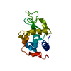
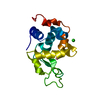
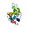
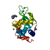
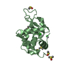
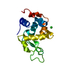
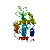
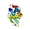
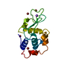
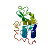
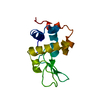
[English] 日本語
Yorodumi
- PDB-6gf0: Lysozyme structure determined from SFX data using a Sheet-on-Shee... -

+
Open data


ID or keywords:

Loading...

-
Basic information

Entry
Database: PDB / ID: 6gf0
TitleLysozyme structure determined from SFX data using a Sheet-on-Sheet chipless chip
ComponentsLysozyme C
KeywordsHYDROLASE / SERIAL CRYSTALLOGRAPHY / FREE-ELECTRON LASER / CHIP CRYSTALLOGRAPHY
Function / homology
Function and homology information


Antimicrobial peptides / Neutrophil degranulation / beta-N-acetylglucosaminidase activity / cell wall macromolecule catabolic process / lysozyme / lysozyme activity / killing of cells of another organism / defense response to Gram-negative bacterium / defense response to Gram-positive bacterium / defense response to bacterium ...Antimicrobial peptides / Neutrophil degranulation / beta-N-acetylglucosaminidase activity / cell wall macromolecule catabolic process / lysozyme / lysozyme activity / killing of cells of another organism / defense response to Gram-negative bacterium / defense response to Gram-positive bacterium / defense response to bacterium / endoplasmic reticulum / extracellular space / identical protein binding / cytoplasm
Similarity search - Function
Lysozyme - #10 / Glycoside hydrolase, family 22, lysozyme / Glycoside hydrolase family 22 domain / Glycosyl hydrolases family 22 (GH22) domain signature. / Glycoside hydrolase, family 22 / C-type lysozyme/alpha-lactalbumin family / Glycosyl hydrolases family 22 (GH22) domain profile. / Alpha-lactalbumin / lysozyme C / Lysozyme / Lysozyme-like domain superfamily ...Lysozyme - #10 / Glycoside hydrolase, family 22, lysozyme / Glycoside hydrolase family 22 domain / Glycosyl hydrolases family 22 (GH22) domain signature. / Glycoside hydrolase, family 22 / C-type lysozyme/alpha-lactalbumin family / Glycosyl hydrolases family 22 (GH22) domain profile. / Alpha-lactalbumin / lysozyme C / Lysozyme / Lysozyme-like domain superfamily / Orthogonal Bundle / Mainly Alpha
Similarity search - Domain/homology
Biological speciesGallus gallus (chicken)
MethodX-RAY DIFFRACTION / FREE ELECTRON LASER / FOURIER SYNTHESIS / Resolution: 2.07 Å
AuthorsDoak, R.B. / Gorel, A. / Foucar, L. / Gruenbein, M.L. / Hilpert, M. / Kloos, M. / Nass Kovacs, G. / Roome, C. / Shoeman, R.L. / Stricker, M. ...Doak, R.B. / Gorel, A. / Foucar, L. / Gruenbein, M.L. / Hilpert, M. / Kloos, M. / Nass Kovacs, G. / Roome, C. / Shoeman, R.L. / Stricker, M. / Tono, K. / You, D. / Ueda, K. / Sherrel, D. / Owen, R. / Barends, T.R.M. / Schlichting, I.
CitationJournal: Acta Crystallogr D Struct Biol / Year: 2018
Title: Crystallography on a chip - without the chip: sheet-on-sheet sandwich.
Authors: Doak, R.B. / Nass Kovacs, G. / Gorel, A. / Foucar, L. / Barends, T.R.M. / Grunbein, M.L. / Hilpert, M. / Kloos, M. / Roome, C.M. / Shoeman, R.L. / Stricker, M. / Tono, K. / You, D. / Ueda, K. ...Authors: Doak, R.B. / Nass Kovacs, G. / Gorel, A. / Foucar, L. / Barends, T.R.M. / Grunbein, M.L. / Hilpert, M. / Kloos, M. / Roome, C.M. / Shoeman, R.L. / Stricker, M. / Tono, K. / You, D. / Ueda, K. / Sherrell, D.A. / Owen, R.L. / Schlichting, I.
History
DepositionApr 27, 2018Deposition site: PDBE / Processing site: PDBE
Revision 1.0Oct 17, 2018Provider: repository / Type: Initial release
Revision 1.1Nov 14, 2018Group: Data collection / Structure summary / Category: diffrn / entity
Item: _diffrn.pdbx_serial_crystal_experiment / _entity.formula_weight
Revision 1.2Jan 17, 2024Group: Data collection / Database references / Refinement description
Category: chem_comp_atom / chem_comp_bond ...chem_comp_atom / chem_comp_bond / database_2 / pdbx_initial_refinement_model
Item: _database_2.pdbx_DOI / _database_2.pdbx_database_accession

-
Structure visualization

Structure viewerMolecule:
MolmilJmol/JSmol

Downloads & links

-
Assembly

Deposited unit
A: Lysozyme C


Theoretical massNumber of molelcules
Total (without water)14,3311
Polymers14,3311
Non-polymers00
Water77543
1


  • Idetical with deposited unit
  • defined by author&software
TypeNameSymmetry operationNumber
identity operation1_555x,y,z1
Buried area0 Å2
ΔGint0 kcal/mol
Surface area6690 Å2
MethodPISA
Unit cell
Length a, b, c (Å)79.100, 79.100, 39.200
Angle α, β, γ (deg.)90.00, 90.00, 90.00
Int Tables number96
Space group name H-MP43212
Components on special symmetry positions
IDModelComponents
11A-213-

HOH

21A-243-

HOH

-
Components

#1: Protein Lysozyme C / 1 / 4-beta-N-acetylmuramidase C / Allergen Gal d IV


Mass: 14331.160 Da / Num. of mol.: 1 / Source method: isolated from a natural source / Source: (natural) Gallus gallus (chicken) / References: UniProt: P00698, lysozyme
#2: Water ChemComp-HOH / water / Water


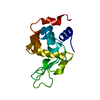
Mass: 18.015 Da / Num. of mol.: 43 / Source method: isolated from a natural source / Formula: H2O

-
Experimental details

-
Experiment

ExperimentMethod: X-RAY DIFFRACTION / Number of used crystals: 1

-
Sample preparation

CrystalDensity Matthews: 2.14 Å3/Da / Density % sol: 42.5 %
Crystal growTemperature: 293 K / Method: batch mode
Details: 20 % NaCl, 6 % PEG 6000, 0.1 M sodium acetate pH 3.0

-
Data collection

DiffractionMean temperature: 293 K / Serial crystal experiment: Y
Diffraction sourceSource: FREE ELECTRON LASER / Site: SACLA / Beamline: BL3 / Wavelength: 1.77 Å
DetectorType: MPCCD / Detector: CCD / Date: Dec 18, 2017
RadiationProtocol: SINGLE WAVELENGTH / Monochromatic (M) / Laue (L): M / Scattering type: x-ray
Radiation wavelengthWavelength: 1.77 Å / Relative weight: 1
ReflectionResolution: 2.07→40 Å / Num. obs: 7417 / % possible obs: 100 % / Redundancy: 283 % / CC1/2: 0.986 / R split: 0.09 / Net I/σ(I): 10.3
Reflection shellResolution: 2.07→2.2 Å / Redundancy: 42 % / Mean I/σ(I) obs: 2.8 / CC1/2: 0.773 / R split: 0.324 / % possible all: 100

-
Processing

Software
NameVersionClassification
REFMAC5.8.0073refinement
CrystFELdata reduction
CrystFELdata scaling
REFMACphasing
RefinementMethod to determine structure: FOURIER SYNTHESIS
Starting model: 4n5r
Resolution: 2.07→39.55 Å / Cor.coef. Fo:Fc: 0.95 / Cor.coef. Fo:Fc free: 0.924 / SU B: 4.616 / SU ML: 0.125 / Cross valid method: THROUGHOUT / ESU R: 0.237 / ESU R Free: 0.186 / Details: HYDROGENS HAVE BEEN ADDED IN THE RIDING POSITIONS
RfactorNum. reflection% reflectionSelection details
Rfree0.23483 385 4.9 %RANDOM
Rwork0.19862 ---
obs0.20034 7417 98.08 %-
Solvent computationIon probe radii: 0.8 Å / Shrinkage radii: 0.8 Å / VDW probe radii: 1.2 Å
Displacement parametersBiso mean: 31.191 Å2
Baniso -1Baniso -2Baniso -3
1--0.37 Å20 Å20 Å2
2---0.37 Å2-0 Å2
3---0.74 Å2
Refinement stepCycle: 1 / Resolution: 2.07→39.55 Å
ProteinNucleic acidLigandSolventTotal
Num. atoms992 0 0 43 1035
Refine LS restraints
Refine-IDTypeDev idealDev ideal targetNumber
X-RAY DIFFRACTIONr_bond_refined_d0.0060.0191016
X-RAY DIFFRACTIONr_bond_other_d0.0010.02929
X-RAY DIFFRACTIONr_angle_refined_deg0.9621.8971376
X-RAY DIFFRACTIONr_angle_other_deg0.79132115
X-RAY DIFFRACTIONr_dihedral_angle_1_deg5.2395127
X-RAY DIFFRACTIONr_dihedral_angle_2_deg33.0882350
X-RAY DIFFRACTIONr_dihedral_angle_3_deg12.61515164
X-RAY DIFFRACTIONr_dihedral_angle_4_deg17.2551511
X-RAY DIFFRACTIONr_chiral_restr0.0530.2142
X-RAY DIFFRACTIONr_gen_planes_refined0.0020.021195
X-RAY DIFFRACTIONr_gen_planes_other0.0010.02270
X-RAY DIFFRACTIONr_nbd_refined
X-RAY DIFFRACTIONr_nbd_other
X-RAY DIFFRACTIONr_nbtor_refined
X-RAY DIFFRACTIONr_nbtor_other
X-RAY DIFFRACTIONr_xyhbond_nbd_refined
X-RAY DIFFRACTIONr_xyhbond_nbd_other
X-RAY DIFFRACTIONr_metal_ion_refined
X-RAY DIFFRACTIONr_metal_ion_other
X-RAY DIFFRACTIONr_symmetry_vdw_refined
X-RAY DIFFRACTIONr_symmetry_vdw_other
X-RAY DIFFRACTIONr_symmetry_hbond_refined
X-RAY DIFFRACTIONr_symmetry_hbond_other
X-RAY DIFFRACTIONr_symmetry_metal_ion_refined
X-RAY DIFFRACTIONr_symmetry_metal_ion_other
X-RAY DIFFRACTIONr_mcbond_it0.8513.005511
X-RAY DIFFRACTIONr_mcbond_other0.853.003510
X-RAY DIFFRACTIONr_mcangle_it1.4584.501637
X-RAY DIFFRACTIONr_mcangle_other1.4574.504638
X-RAY DIFFRACTIONr_scbond_it1.0893.188505
X-RAY DIFFRACTIONr_scbond_other1.0883.193506
X-RAY DIFFRACTIONr_scangle_it
X-RAY DIFFRACTIONr_scangle_other1.8984.716740
X-RAY DIFFRACTIONr_long_range_B_refined3.21724.2841199
X-RAY DIFFRACTIONr_long_range_B_other3.21524.2831195
X-RAY DIFFRACTIONr_rigid_bond_restr
X-RAY DIFFRACTIONr_sphericity_free
X-RAY DIFFRACTIONr_sphericity_bonded
LS refinement shellResolution: 2.074→2.128 Å / Total num. of bins used: 20
RfactorNum. reflection% reflection
Rfree0.32 12 -
Rwork0.45 410 -
obs--73.91 %

+
About Yorodumi

-
News

-
Feb 9, 2022. New format data for meta-information of EMDB entries

New format data for meta-information of EMDB entries

  • Version 3 of the EMDB header file is now the official format.
  • The previous official version 1.9 will be removed from the archive.

Related info.:EMDB header

External links:wwPDB to switch to version 3 of the EMDB data model

-
Aug 12, 2020. Covid-19 info

Covid-19 info

URL: https://pdbj.org/emnavi/covid19.php

New page: Covid-19 featured information page in EM Navigator.

Related info.:Covid-19 info / Mar 5, 2020. Novel coronavirus structure data

+
Mar 5, 2020. Novel coronavirus structure data

Novel coronavirus structure data

Related info.:Yorodumi Speices / Aug 12, 2020. Covid-19 info

External links:COVID-19 featured content - PDBj / Molecule of the Month (242):Coronavirus Proteases

+
Jan 31, 2019. EMDB accession codes are about to change! (news from PDBe EMDB page)

EMDB accession codes are about to change! (news from PDBe EMDB page)

  • The allocation of 4 digits for EMDB accession codes will soon come to an end. Whilst these codes will remain in use, new EMDB accession codes will include an additional digit and will expand incrementally as the available range of codes is exhausted. The current 4-digit format prefixed with “EMD-” (i.e. EMD-XXXX) will advance to a 5-digit format (i.e. EMD-XXXXX), and so on. It is currently estimated that the 4-digit codes will be depleted around Spring 2019, at which point the 5-digit format will come into force.
  • The EM Navigator/Yorodumi systems omit the EMD- prefix.

Related info.:Q: What is EMD? / ID/Accession-code notation in Yorodumi/EM Navigator

External links:EMDB Accession Codes are Changing Soon! / Contact to PDBj

+
Jul 12, 2017. Major update of PDB

Major update of PDB

  • wwPDB released updated PDB data conforming to the new PDBx/mmCIF dictionary.
  • This is a major update changing the version number from 4 to 5, and with Remediation, in which all the entries are updated.
  • In this update, many items about electron microscopy experimental information are reorganized (e.g. em_software).
  • Now, EM Navigator and Yorodumi are based on the updated data.

External links:wwPDB Remediation / Enriched Model Files Conforming to OneDep Data Standards Now Available in the PDB FTP Archive

-
Yorodumi

Thousand views of thousand structures

  • Yorodumi is a browser for structure data from EMDB, PDB, SASBDB, etc.
  • This page is also the successor to EM Navigator detail page, and also detail information page/front-end page for Omokage search.
  • The word "yorodu" (or yorozu) is an old Japanese word meaning "ten thousand". "mi" (miru) is to see.

Related info.:EMDB / PDB / SASBDB / Comparison of 3 databanks / Yorodumi Search / Aug 31, 2016. New EM Navigator & Yorodumi / Yorodumi Papers / Jmol/JSmol / Function and homology information / Changes in new EM Navigator and Yorodumi

Read more