Mass: 18.015 Da / Num. of mol.: 59 / Source method: isolated from a natural source / Formula: H2O
-
Experimental details
-
Experiment
Experiment
Method
Number of used crystals
X-RAY DIFFRACTION
1
NEUTRON DIFFRACTION
-
Sample preparation
Crystal
Density Matthews: 2.19 Å3/Da / Density % sol: 44 %
Crystal grow
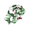
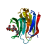
Temperature: 293 K / Method: vapor diffusion, sitting drop / pH: 7.5 Details: 12-15% PEG 4000 OR PEG 3000, 0.1M MGCL2, 0.015M BETA MERCAPTOETHANOL, 0.1M TRIS-DCL, PD 7.9, 0.4M NASCN. ALL DISSOLVED IN D2O. CRYSTAL GROWN IN A 15 + 15 MICROLITRE SITTING DROP THAT WAS ...Details: 12-15% PEG 4000 OR PEG 3000, 0.1M MGCL2, 0.015M BETA MERCAPTOETHANOL, 0.1M TRIS-DCL, PD 7.9, 0.4M NASCN. ALL DISSOLVED IN D2O. CRYSTAL GROWN IN A 15 + 15 MICROLITRE SITTING DROP THAT WAS FIRST EQUILIBRATED FOR 1 WEEK. A SMALL CRYSTAL GROWN AT 20-28% PEG WAS THEN INTRODUCED. THE DROP WAS FED WITH FRESH PROTEIN BY ADDING 3-4 MICROLITRES OF PROTEIN WITH 10 MM LACTOSE EVERY 3-4 DAYS FOR 3 MONTHS. LACTOSE WAS THEN REMOVED IN TWO STEPS, FIRST BY DIALYSING AGAINST 1M GLYCEROL FOR 1 MONTH THEN AGAINST BUFFER WITH NO GLYCEROL FOR 1 MONTH. FOR DETAILS, SEE MANZONI ET AL. (2016).
-
Data collection
Diffraction
ID
Mean temperature (K)
Crystal-ID
1
100
1
2
298
1
Diffraction source
Source
Site
Beamline
ID
Wavelength (Å)
SYNCHROTRON
ESRF
BM30A
1
0.98081
NUCLEAR REACTOR
ILL
LADIIII
2
3.35-4.35
Detector
Type
ID
Detector
Date
Details
ADSC QUANTUM 315r
1
CCD
Mar 10, 2017
FOCUSINGMIRRORS
2
IMAGE PLATE
Feb 15, 2017
Radiation
ID
Monochromator
Protocol
Monochromatic (M) / Laue (L)
Scattering type
Wavelength-ID
1
DOUBLECRYSTAL
SINGLEWAVELENGTH
M
x-ray
1
2
LAUE
L
neutron
2
Radiation wavelength
ID
Wavelength (Å)
Relative weight
1
0.98081
1
2
3.35
1
3
4.35
1
Reflection
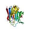
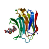
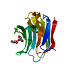
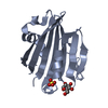
Entry-ID: 6F2Q
Resolution (Å)
Num. obs
% possible obs (%)
Redundancy (%)
CC1/2
Rmerge(I) obs
Rpim(I) all
Diffraction-ID
Net I/σ(I)
Rsym value
1.03-28
70173
99.8
14
1
0.044
0.012
1
35.5
1.8-30
9844
83.8
4.9
0.993
0.053
2
8.5
0.132
Reflection shell
Resolution (Å)
Redundancy (%)
Rmerge(I) obs
Mean I/σ(I) obs
Num. unique obs
CC1/2
Rpim(I) all
Diffraction-ID
% possible all
1.03-1.06
9.3
2.357
1
5138
0.355
0.933
1
99.2
1.8-1.9
3.4
0.194
1177
0.917
0.091
2
67.1
-
Processing
Software
Name
Version
Classification
PHENIX
(1.12_2829: ???)
refinement
XDS
datareduction
LAUEGEN
datascaling
PHENIX
phasing
Refinement
SU ML: 0.11 / Cross valid method: THROUGHOUT / Method to determine structure: FOURIER SYNTHESIS / Phase error: 13.66 / Shrinkage radii: 0.9 Å / VDW probe radii: 1.11 Å / Starting model: 6EYM
In the structure databanks used in Yorodumi, some data are registered as the other names, "COVID-19 virus" and "2019-nCoV". Here are the details of the virus and the list of structure data.
Jan 31, 2019. EMDB accession codes are about to change! (news from PDBe EMDB page)
EMDB accession codes are about to change! (news from PDBe EMDB page)
The allocation of 4 digits for EMDB accession codes will soon come to an end. Whilst these codes will remain in use, new EMDB accession codes will include an additional digit and will expand incrementally as the available range of codes is exhausted. The current 4-digit format prefixed with “EMD-” (i.e. EMD-XXXX) will advance to a 5-digit format (i.e. EMD-XXXXX), and so on. It is currently estimated that the 4-digit codes will be depleted around Spring 2019, at which point the 5-digit format will come into force.
The EM Navigator/Yorodumi systems omit the EMD- prefix.
Related info.:Q: What is EMD? / ID/Accession-code notation in Yorodumi/EM Navigator
Yorodumi is a browser for structure data from EMDB, PDB, SASBDB, etc.
This page is also the successor to EM Navigator detail page, and also detail information page/front-end page for Omokage search.
The word "yorodu" (or yorozu) is an old Japanese word meaning "ten thousand". "mi" (miru) is to see.
Related info.:EMDB / PDB / SASBDB / Comparison of 3 databanks / Yorodumi Search / Aug 31, 2016. New EM Navigator & Yorodumi / Yorodumi Papers / Jmol/JSmol / Function and homology information / Changes in new EM Navigator and Yorodumi
 Movie
Movie Controller

 Yorodumi
 Open data
 Basic information
 Components
 Keywords
 Function and homology information
 Homo sapiens (human)
 X-RAY DIFFRACTION / NEUTRON DIFFRACTION /
  Authors
 Sweden, 1items
  Citation
 Structure visualization
 Downloads & links
 Other downloads
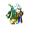


 PDBj
PDBj


 Assembly
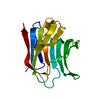

 Mass: 18.015 Da / Num. of mol.: 59 / Source method: isolated from a natural source / Formula: H2O
 Sample preparation
 Processing