 Movie
Movie Controller


+ Open data

- Basic information
| Entry | Database: PDB / ID: 6bim | ||||||
|---|---|---|---|---|---|---|---|
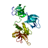
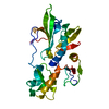
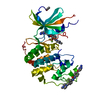
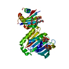
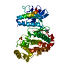
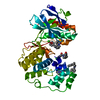
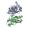
| Title | Structure of NlpC1 from Trichomonas vaginalis | ||||||
 Components Components | Clan CA, family C40, NlpC/P60 superfamily cysteine peptidase Papain-like protease Papain-like protease | ||||||
 Keywords Keywords | HYDROLASE / peptidoglycan / peptidase / NlpC protein | ||||||
| Function / homology | Bacterial SH3 domain / NlpC/P60 domain profile. / SH3-like domain, bacterial-type / Endopeptidase, NLPC/P60 domain / NlpC/P60 family / Papain-like cysteine peptidase superfamily / Clan CA, family C40, NlpC/P60 superfamily cysteine peptidase Function and homology information Function and homology information | ||||||
| Biological species |  Trichomonas vaginalis (eukaryote) Trichomonas vaginalis (eukaryote) | ||||||
| Method | X-RAY DIFFRACTION / SIR / Resolution: 1.547 Å | ||||||
 Authors Authors | Pinheiro, J. / Simoes-Barbosa, A. / Goldstone, D.C. | ||||||
 Citation Citation | Journal: Mbio / Year: 2018 Title: The ProtozoanTrichomonas vaginalisTargets Bacteria with Laterally Acquired NlpC/P60 Peptidoglycan Hydrolases. Authors: Pinheiro, J. / Biboy, J. / Vollmer, W. / Hirt, R.P. / Keown, J.R. / Artuyants, A. / Black, M.M. / Goldstone, D.C. / Simoes-Barbosa, A. | ||||||
| History |
|
- Structure visualization
| Structure viewer | Molecule: MolmilJmol/JSmol |
|---|
- Downloads & links
-Download
| PDBx/mmCIF format | 6bim.cif.gz | 124.9 KB | Display | PDBx/mmCIF format |
|---|---|---|---|---|
| PDB format | pdb6bim.ent.gz | 103.8 KB | Display | PDB format |
| PDBx/mmJSON format | 6bim.json.gz | Tree view | PDBx/mmJSON format | |
| Others |  Other downloads Other downloads |
-Validation report
| Arichive directory | https://data.pdbj.org/pub/pdb/validation_reports/bi/6bimftp://data.pdbj.org/pub/pdb/validation_reports/bi/6bim | HTTPS FTP |
|---|
-Related structure data


-Links
 PDBj
PDBj- Assembly
| Deposited unit | 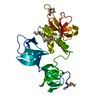
| ||||||||
|---|---|---|---|---|---|---|---|---|---|
| 1 |
| ||||||||
| Unit cell |
| ||||||||
| Components on special symmetry positions |
|
-Components
| #1: Protein | Papain-like protease Mass: 31937.910 Da / Num. of mol.: 1 Source method: isolated from a genetically manipulated source Source: (gene. exp.) Trichomonas vaginalis (eukaryote) / Gene: TVAG_119910 / Production host:  Escherichia coli (E. coli) / Strain (production host): BL21(DE3) / References: UniProt: A2D7D7 Escherichia coli (E. coli) / Strain (production host): BL21(DE3) / References: UniProt: A2D7D7 |
|---|---|
| #2: Water | ChemComp-HOH / Water Mass: 18.015 Da / Num. of mol.: 516 / Source method: isolated from a natural source / Formula: H2O Mass: 18.015 Da / Num. of mol.: 516 / Source method: isolated from a natural source / Formula: H2O |
-Experimental details
-Experiment
| Experiment | Method: X-RAY DIFFRACTION / Number of used crystals: 1 |
|---|
- Sample preparation
| Crystal | Density Matthews: 2.33 Å3/Da / Density % sol: 47.18 % |
|---|---|
| Crystal grow | Temperature: 291 K / Method: vapor diffusion, sitting drop / Details: Morpheus screen A1 |
-Data collection
| Diffraction | Mean temperature: 110 K |
|---|---|
| Diffraction source | Source: ROTATING ANODE / Type: RIGAKU MICROMAX-007 HF / Wavelength: 1.5418 Å |
| Detector | Type: MAR scanner 345 mm plate / Detector: IMAGE PLATE / Date: Jan 1, 2015 |
| Radiation | Protocol: SINGLE WAVELENGTH / Monochromatic (M) / Laue (L): M / Scattering type: x-ray |
| Radiation wavelength | Wavelength: 1.5418 Å / Relative weight: 1 |
| Reflection | Resolution: 1.54→29.24 Å / Num. obs: 83416 / % possible obs: 98.8 % / Redundancy: 5.7 % / Rpim(I) all: 0.03 / Net I/σ(I): 14.1 |
| Reflection shell | Resolution: 1.54→1.57 Å / Redundancy: 5 % / Mean I/σ(I) obs: 4.1 / Num. unique obs: 1744 / Rpim(I) all: 0.112 / % possible all: 81 |
- Processing
| Software |
| ||||||||||||||||||||||||||||||||||||||||||||||||||||||||||||||||||||||||||||||||||||||||||||||||||||||||||||||||||||||||||||||||||||||||||||||||||||||||||||||||||||||||||||||||||||||||||||||||||||||||||||||||||
|---|---|---|---|---|---|---|---|---|---|---|---|---|---|---|---|---|---|---|---|---|---|---|---|---|---|---|---|---|---|---|---|---|---|---|---|---|---|---|---|---|---|---|---|---|---|---|---|---|---|---|---|---|---|---|---|---|---|---|---|---|---|---|---|---|---|---|---|---|---|---|---|---|---|---|---|---|---|---|---|---|---|---|---|---|---|---|---|---|---|---|---|---|---|---|---|---|---|---|---|---|---|---|---|---|---|---|---|---|---|---|---|---|---|---|---|---|---|---|---|---|---|---|---|---|---|---|---|---|---|---|---|---|---|---|---|---|---|---|---|---|---|---|---|---|---|---|---|---|---|---|---|---|---|---|---|---|---|---|---|---|---|---|---|---|---|---|---|---|---|---|---|---|---|---|---|---|---|---|---|---|---|---|---|---|---|---|---|---|---|---|---|---|---|---|---|---|---|---|---|---|---|---|---|---|---|---|---|---|---|---|---|
| Refinement | Method to determine structure: SIR / Resolution: 1.547→29.059 Å / SU ML: 0.14 / Cross valid method: FREE R-VALUE / σ(F): 1.36 / Phase error: 15.67
| ||||||||||||||||||||||||||||||||||||||||||||||||||||||||||||||||||||||||||||||||||||||||||||||||||||||||||||||||||||||||||||||||||||||||||||||||||||||||||||||||||||||||||||||||||||||||||||||||||||||||||||||||||
| Solvent computation | Shrinkage radii: 0.9 Å / VDW probe radii: 1.11 Å | ||||||||||||||||||||||||||||||||||||||||||||||||||||||||||||||||||||||||||||||||||||||||||||||||||||||||||||||||||||||||||||||||||||||||||||||||||||||||||||||||||||||||||||||||||||||||||||||||||||||||||||||||||
| Refinement step | Cycle: LAST / Resolution: 1.547→29.059 Å
| ||||||||||||||||||||||||||||||||||||||||||||||||||||||||||||||||||||||||||||||||||||||||||||||||||||||||||||||||||||||||||||||||||||||||||||||||||||||||||||||||||||||||||||||||||||||||||||||||||||||||||||||||||
| Refine LS restraints |
| ||||||||||||||||||||||||||||||||||||||||||||||||||||||||||||||||||||||||||||||||||||||||||||||||||||||||||||||||||||||||||||||||||||||||||||||||||||||||||||||||||||||||||||||||||||||||||||||||||||||||||||||||||
| LS refinement shell |
|
