negative regulation of bone trabecula formation / Vitamin D (calciferol) metabolism / dense fibrillar component / SUMOylation of intracellular receptors / Nuclear Receptor transcription pathway / bile acid nuclear receptor activity / response to bile acid / positive regulation of parathyroid hormone secretion / phosphate ion transmembrane transport / apoptotic process involved in mammary gland involution ...negative regulation of bone trabecula formation / Vitamin D (calciferol) metabolism / dense fibrillar component / SUMOylation of intracellular receptors / Nuclear Receptor transcription pathway / bile acid nuclear receptor activity / response to bile acid / positive regulation of parathyroid hormone secretion / phosphate ion transmembrane transport / apoptotic process involved in mammary gland involution / vitamin D binding / cellular response to vitamin D / positive regulation of apoptotic process involved in mammary gland involution / calcitriol binding / lithocholic acid binding / positive regulation of keratinocyte differentiation / negative regulation of ossification / positive regulation of vitamin D receptor signaling pathway / vitamin D receptor signaling pathway / bile acid signaling pathway / intestinal absorption / response to aldosterone / RNA polymerase II intronic transcription regulatory region sequence-specific DNA binding / locomotor rhythm / mammary gland branching involved in pregnancy / regulation of calcium ion transport / aryl hydrocarbon receptor binding / decidualization / regulation of lipid metabolic process / cellular response to Thyroglobulin triiodothyronine / regulation of glucose metabolic process / Synthesis of bile acids and bile salts / negative regulation of keratinocyte proliferation / Endogenous sterols / Synthesis of bile acids and bile salts via 27-hydroxycholesterol / heterochromatin / Synthesis of bile acids and bile salts via 7alpha-hydroxycholesterol / nuclear retinoid X receptor binding / Recycling of bile acids and salts / regulation of cellular response to insulin stimulus / cellular response to hormone stimulus / positive regulation of adipose tissue development / T-tubule / peroxisome proliferator activated receptor signaling pathway / RORA activates gene expression / lactation / Regulation of lipid metabolism by PPARalpha / BMAL1:CLOCK,NPAS2 activates circadian gene expression / SUMOylation of transcription cofactors / nuclear receptor coactivator activity / Activation of gene expression by SREBF (SREBP) / response to progesterone / skeletal system development / apoptotic signaling pathway / nuclear receptor binding / animal organ morphogenesis / circadian regulation of gene expression / euchromatin / Heme signaling / mRNA transcription by RNA polymerase II / Activated PKN1 stimulates transcription of AR (androgen receptor) regulated genes KLK2 and KLK3 / cell morphogenesis / Transcriptional activation of mitochondrial biogenesis / intracellular calcium ion homeostasis / PPARA activates gene expression / Cytoprotection by HMOX1 / nuclear matrix / Transcriptional regulation of white adipocyte differentiation / response to calcium ion / RNA polymerase II transcription regulator complex / cellular response to amyloid-beta / nuclear receptor activity / calcium ion transport / Circadian Clock / response to estradiol / heart development / HATs acetylate histones / Estrogen-dependent gene expression / transcription regulator complex / sequence-specific DNA binding / cell differentiation / transcription coactivator activity / protein dimerization activity / nuclear body / receptor complex / RNA polymerase II cis-regulatory region sequence-specific DNA binding / DNA-binding transcription factor activity / protein domain specific binding / negative regulation of cell population proliferation / negative regulation of DNA-templated transcription / chromatin binding / chromatin / positive regulation of gene expression / regulation of DNA-templated transcription / regulation of transcription by RNA polymerase II / perinuclear region of cytoplasm / negative regulation of transcription by RNA polymerase II / positive regulation of transcription by RNA polymerase II / protein-containing complex / DNA binding Similarity search - Function
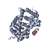
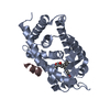
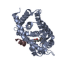
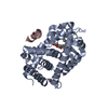
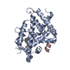
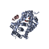
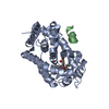
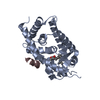
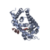
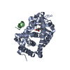
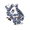
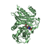
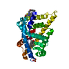
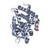
Journal: J. Med. Chem. / Year: 2017 Title: Vitamin D Analogues with a p-Hydroxyphenyl Group at the C25 Position: Crystal Structure of Vitamin D Receptor Ligand-Binding Domain Complexed with the Ligand Explains the Mechanism Underlying ...Title: Vitamin D Analogues with a p-Hydroxyphenyl Group at the C25 Position: Crystal Structure of Vitamin D Receptor Ligand-Binding Domain Complexed with the Ligand Explains the Mechanism Underlying Full Antagonistic Action Authors: Kato, A. / Yamao, M. / Hashihara, Y. / Ishida, H. / Itoh, T. / Yamamoto, K.
 Movie
Movie Controller

 Yorodumi
 Open data
 Basic information
 Components
 Keywords
 TRANSCRIPTION /
  Function and homology information

 Authors
 Citation
 Structure visualization
 Downloads & links
 Other downloads
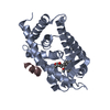


 PDBj
PDBj


 Assembly
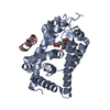

 Mass: 534.769 Da / Num. of mol.: 1 / Source method: obtained synthetically / Formula: C35H50O4
 Mass: 18.015 Da / Num. of mol.: 91 / Source method: isolated from a natural source / Formula: H2O
 Sample preparation
 / Beamline: BL-17A / Wavelength: 1 Å
 Processing